Sunday Poster Session
Category: Liver
P1006 - Small Bowel Variceal Hemorrhage Requiring TIPS: An Inconspicuous Etiology of Melena and Anemia in Patients with Cirrhosis
Sunday, October 22, 2023
3:30 PM - 7:00 PM PT
Location: Exhibit Hall

Has Audio
.jpg)
Albert Manudhane, MD
The Ohio State University Wexner Medical Center
Columbus, OH
Presenting Author(s)
Albert Manudhane, MD1, Madalina Butnariu, MD1, Lindsay A. Sobotka, DO2
1The Ohio State University, Columbus, OH; 2The Ohio State Wexner Medical Center, Columbus, OH
Introduction: Patients with cirrhosis can develop decompensated disease characterized by sequela of portal hypertension including ascites, hepatic encephalopathy, and variceal hemorrhage. Variceal hemorrhage is typically a result of esophagogastric or rectal varices but rarely can be due to small bowel varices. Small bowel varices almost exclusively occurs in patients that also have esophagogastric varices and are noted in less the 3% of patients with cirrhosis without evidence of portal hypertension that underwent video capsule endoscopy (VCE). We describe a case of obscure bleeding due to isolated small bowel varices leading to life-threatening gastrointestinal bleed that was treated with transjugular portosystemic intrahepatic shunt (TIPS).
Case Description/Methods: A 45-year-old female with a history of compensated treatment naïve hepatitis C cirrhosis presented for melena. Labs were notable for a hemoglobin 2.4 g/dL, platelets 225 x 109/L, creatinine 1.22mg/dL, sodium 139mmol/L, total bilirubin 0.6 mg/dL, and INR 1.4. Her MELD-Na score was 12. CT abdomen pelvis revealed cirrhosis and splenomegaly. EGD showed LA Grade A esophagitis but no esophagogastric varices or portal hypertensive gastropathy. Colonoscopy displayed a 6mm cecal polyp and hemorrhiods but no obvious source of her melena and severe anemia. VCE demonstrated a distal ileum submucosal lesion with stigmata of recent bleeding concerning for small bowel varices. CT enterography showed a small cluster of prominent collateral veins in the ileum consistent with small bowel varices. She underwent a TIPS where an 8-10mm covered stent was placed resulting in a reduction of the HVPG to 6 mmHg from 12mmHg. After the TIPS, her hemoglobin remained stable with no overt bleeding.
Discussion: This case demonstrates the need to evaluate for small bowel varices in patients with cirrhosis who present with obscure gastrointestinal bleeding, even in individuals without esophagogastric varices or portal hypertensive gastropathy. VCE is useful as it has the potential to other etiologies of bleeding including small bowel ulcers, erosions, and angioectasias in addition to varices. CT Angiogram or enterography can confirm the presence of small bowel varices. Management includes TIPS, interventional radiology embolization, cyanoacrylate injection, or surgical resection of effected small bowel. In summary, the diagnosis of small bowel variceal hemorrhage should be considered in any patient with cirrhosis, regardless of known sequela of portal hypertension.

Disclosures:
Albert Manudhane, MD1, Madalina Butnariu, MD1, Lindsay A. Sobotka, DO2. P1006 - Small Bowel Variceal Hemorrhage Requiring TIPS: An Inconspicuous Etiology of Melena and Anemia in Patients with Cirrhosis, ACG 2023 Annual Scientific Meeting Abstracts. Vancouver, BC, Canada: American College of Gastroenterology.
1The Ohio State University, Columbus, OH; 2The Ohio State Wexner Medical Center, Columbus, OH
Introduction: Patients with cirrhosis can develop decompensated disease characterized by sequela of portal hypertension including ascites, hepatic encephalopathy, and variceal hemorrhage. Variceal hemorrhage is typically a result of esophagogastric or rectal varices but rarely can be due to small bowel varices. Small bowel varices almost exclusively occurs in patients that also have esophagogastric varices and are noted in less the 3% of patients with cirrhosis without evidence of portal hypertension that underwent video capsule endoscopy (VCE). We describe a case of obscure bleeding due to isolated small bowel varices leading to life-threatening gastrointestinal bleed that was treated with transjugular portosystemic intrahepatic shunt (TIPS).
Case Description/Methods: A 45-year-old female with a history of compensated treatment naïve hepatitis C cirrhosis presented for melena. Labs were notable for a hemoglobin 2.4 g/dL, platelets 225 x 109/L, creatinine 1.22mg/dL, sodium 139mmol/L, total bilirubin 0.6 mg/dL, and INR 1.4. Her MELD-Na score was 12. CT abdomen pelvis revealed cirrhosis and splenomegaly. EGD showed LA Grade A esophagitis but no esophagogastric varices or portal hypertensive gastropathy. Colonoscopy displayed a 6mm cecal polyp and hemorrhiods but no obvious source of her melena and severe anemia. VCE demonstrated a distal ileum submucosal lesion with stigmata of recent bleeding concerning for small bowel varices. CT enterography showed a small cluster of prominent collateral veins in the ileum consistent with small bowel varices. She underwent a TIPS where an 8-10mm covered stent was placed resulting in a reduction of the HVPG to 6 mmHg from 12mmHg. After the TIPS, her hemoglobin remained stable with no overt bleeding.
Discussion: This case demonstrates the need to evaluate for small bowel varices in patients with cirrhosis who present with obscure gastrointestinal bleeding, even in individuals without esophagogastric varices or portal hypertensive gastropathy. VCE is useful as it has the potential to other etiologies of bleeding including small bowel ulcers, erosions, and angioectasias in addition to varices. CT Angiogram or enterography can confirm the presence of small bowel varices. Management includes TIPS, interventional radiology embolization, cyanoacrylate injection, or surgical resection of effected small bowel. In summary, the diagnosis of small bowel variceal hemorrhage should be considered in any patient with cirrhosis, regardless of known sequela of portal hypertension.

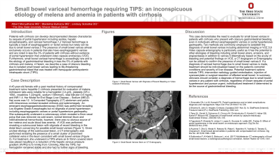
Figure: Figure 1A: Small Bowel Varices with Stigmata of Recent Bleeding on Video Capsule Enterography, and Figure 1B: CT Enterography Findings of Small Bowel Varices
Disclosures:
Albert Manudhane indicated no relevant financial relationships.
Madalina Butnariu indicated no relevant financial relationships.
Lindsay Sobotka indicated no relevant financial relationships.
Albert Manudhane, MD1, Madalina Butnariu, MD1, Lindsay A. Sobotka, DO2. P1006 - Small Bowel Variceal Hemorrhage Requiring TIPS: An Inconspicuous Etiology of Melena and Anemia in Patients with Cirrhosis, ACG 2023 Annual Scientific Meeting Abstracts. Vancouver, BC, Canada: American College of Gastroenterology.
