Sunday Poster Session
Category: Liver
P1025 - Incidental Finding of Hepatic Inflammatory Pseudotumor Immunoglobulin G4-Related Disease With Underlying Chronic Hepatitis C
Sunday, October 22, 2023
3:30 PM - 7:00 PM PT
Location: Exhibit Hall

Has Audio

Malique Delbrune, BS
University of Minnesota
Minneapolis, MN
Presenting Author(s)
Malique Delbrune, BS, Nicha Wongjarupong, MD, Elizabeth S. Aby, MD, Carlos Iwamoto, MD, Mohamed A. Hassan, MD
University of Minnesota, Minneapolis, MN
Introduction: Immunoglobulin G4-related disease (IgG4-RD) is a unique immunological disease that can impact multiple organs. This inflammatory condition can present within the hepatobiliary system with the formation of a hepatic inflammatory pseudotumor (IPT). We present a case of IgG4-RD in a patient with a history of chronic viral hepatitis C infection and a summary of the literature of IgG4-RD presenting in the liver.
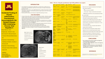
Case Description/Methods: A 67-year-old male with a history of chronic viral hepatitis C infection with a sustained virologic response without advanced hepatic fibrosis presented with a bile duct injury following laparoscopic cholecystectomy for acute cholecystitis. His post operative course was complicated by multiple instances in which percutaneous drains placed became dislodged requiring repositioning. Magnetic resonance imaging (MRI) for drain assessment found an enlarging arterially enhancing lesion involving hepatic segment 3, measuring 2 cm, concerning for hepatocellular carcinoma (HCC) [Figure 1]. Core needle biopsy of the liver was performed and showed an inflammatory process composed of abundant lymphoplasmacytic inflammation with focal storiform type fibrosis. Immunohistochemical stains for IgG was performed and showed markedly increased IgG4 positive plasma cells ( > 50/HPF), which was consistent with IgG4-RD. The additional IgG4 staining was obtained on the gallbladder pathology which was negative for IgG4 positive plasma cell. He was treated with prednisone for three months and mass like structure was not visualized in hepatic segment 3 on the follow up magnetic resonance cholangiopancreatography (MRCP) [Figure 2].
Discussion: A literature search via PubMed using -IgG4, -hepatic, and -pseudotumor found 19 previously published case reports. A common factor discussed amongst cases was that imaging findings were inconclusive in determining if the etiology was malignant or infectious. Interestingly, in cases where quantitative IgG4 serum was reported, 70% did not meet the diagnostic requirement to make the diagnosis of IgG4-RD. There were no other case reports found involving a patient with hepatic pseudotumor from IgG4-RD and a history of chronic viral hepatitis C infection. In our patient the pseudotumor was found incidentally and was treated successfully with a steroid.

Disclosures:
Malique Delbrune, BS, Nicha Wongjarupong, MD, Elizabeth S. Aby, MD, Carlos Iwamoto, MD, Mohamed A. Hassan, MD. P1025 - Incidental Finding of Hepatic Inflammatory Pseudotumor Immunoglobulin G4-Related Disease With Underlying Chronic Hepatitis C, ACG 2023 Annual Scientific Meeting Abstracts. Vancouver, BC, Canada: American College of Gastroenterology.
University of Minnesota, Minneapolis, MN
Introduction: Immunoglobulin G4-related disease (IgG4-RD) is a unique immunological disease that can impact multiple organs. This inflammatory condition can present within the hepatobiliary system with the formation of a hepatic inflammatory pseudotumor (IPT). We present a case of IgG4-RD in a patient with a history of chronic viral hepatitis C infection and a summary of the literature of IgG4-RD presenting in the liver.
Case Description/Methods: A 67-year-old male with a history of chronic viral hepatitis C infection with a sustained virologic response without advanced hepatic fibrosis presented with a bile duct injury following laparoscopic cholecystectomy for acute cholecystitis. His post operative course was complicated by multiple instances in which percutaneous drains placed became dislodged requiring repositioning. Magnetic resonance imaging (MRI) for drain assessment found an enlarging arterially enhancing lesion involving hepatic segment 3, measuring 2 cm, concerning for hepatocellular carcinoma (HCC) [Figure 1]. Core needle biopsy of the liver was performed and showed an inflammatory process composed of abundant lymphoplasmacytic inflammation with focal storiform type fibrosis. Immunohistochemical stains for IgG was performed and showed markedly increased IgG4 positive plasma cells ( > 50/HPF), which was consistent with IgG4-RD. The additional IgG4 staining was obtained on the gallbladder pathology which was negative for IgG4 positive plasma cell. He was treated with prednisone for three months and mass like structure was not visualized in hepatic segment 3 on the follow up magnetic resonance cholangiopancreatography (MRCP) [Figure 2].
Discussion: A literature search via PubMed using -IgG4, -hepatic, and -pseudotumor found 19 previously published case reports. A common factor discussed amongst cases was that imaging findings were inconclusive in determining if the etiology was malignant or infectious. Interestingly, in cases where quantitative IgG4 serum was reported, 70% did not meet the diagnostic requirement to make the diagnosis of IgG4-RD. There were no other case reports found involving a patient with hepatic pseudotumor from IgG4-RD and a history of chronic viral hepatitis C infection. In our patient the pseudotumor was found incidentally and was treated successfully with a steroid.

Figure: Figure 1A. Magnetic resonance imaging (MRI) revealed arterially enhancing lesion arising exophytically in segment III of the liver with associated washout and pseudocapsule.
Figure 1B. Magnetic resonance cholangiopancreatography (MRCP) demonstrating lack of visualization of previously noted lesion in segment III of the liver.
Figure 1B. Magnetic resonance cholangiopancreatography (MRCP) demonstrating lack of visualization of previously noted lesion in segment III of the liver.
Disclosures:
Malique Delbrune indicated no relevant financial relationships.
Nicha Wongjarupong indicated no relevant financial relationships.
Elizabeth Aby indicated no relevant financial relationships.
Carlos Iwamoto indicated no relevant financial relationships.
Mohamed Hassan indicated no relevant financial relationships.
Malique Delbrune, BS, Nicha Wongjarupong, MD, Elizabeth S. Aby, MD, Carlos Iwamoto, MD, Mohamed A. Hassan, MD. P1025 - Incidental Finding of Hepatic Inflammatory Pseudotumor Immunoglobulin G4-Related Disease With Underlying Chronic Hepatitis C, ACG 2023 Annual Scientific Meeting Abstracts. Vancouver, BC, Canada: American College of Gastroenterology.
