Sunday Poster Session
Category: Liver
P1031 - Non-HFE Hemochromatosis Presenting With Intermittent Elevation of Liver Enzymes
Sunday, October 22, 2023
3:30 PM - 7:00 PM PT
Location: Exhibit Hall

Has Audio
- PY
Pradeep Yarra, MD, MSc
Saint Louis University Hospital
St. Louis, MO
Presenting Author(s)
Kishore Karri, MD1, Pradeep Yarra, MD, MSc2, Manar Shahwan, MD3, Nimish Thakral, MD1
1University of Kentucky, Lexington, KY; 2Saint Louis University Hospital, St. Louis, MO; 3Saint Louis University, St. Louis, MO
Introduction: Hereditary Hemochromatosis (HH) is an autosomal recessive disorder characterized by abnormally high levels of intestinal iron absorption leading to iron overload in organs and classically associated homozygous mutations in HFE (human factors engineering) gene. We present a case of an elderly african american man evaluated for elevated liver enzymes.
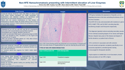
Case Description/Methods: 65-year-old male with past medical history status post living donor kidney transplant for end stage renal disease secondary to hypertension and diabetes mellitus on tacrolimus and prednisone for intermittent elevation of liver enzymes since last five years. Patient did not receive transfusions or has history of thalassemia or alcohol intake. No family history of HH is first or second-degree relatives. Patient denied any symptoms or signs of chronic liver disease. Review of liver function tests revealed normal total bilirubin, albumin, prothrombin time, alkaline phosphatase but elevated alanine transaminase (ALT) at 147 U/L and aspartate transaminase (AST) 96 U/L. Complete blood count showed normal hemoglobin at 13.8 g/dL with MCV at 96.3 fL. Review of records show intermittently elevated AST and ALT for last five years, but more persistent in last year. Ferritin was elevated at 2,983 ng/mL with previous elevations more than 3500. Transferrin saturation was elevated at 56 %, transferrin at 163 mg/dL and normal iron at 114 ug/dL. Anti-nuclear antibodies were detected, showed speckled pattern at 1:160 titers. Other work up for chronic liver disease including A1AT, ceruloplasmin, immunoglobulins, Hepatitis panel, celiac and autoimmune liver diseases work up were negative. Ultrasound abdomen showed mild increased echogenicity. Fibro scan revealed low probability of advanced fibrosis (6.6 kPa) with mild steatosis. HH mutation panel was negative for C282Y, H63D, S65C mutations. Liver biopsy done showed 3+ hepatocellular iron and prominent kupffer cell/portal macrophage iron with no fibrosis confirming the diagnosis of non-HFE hemochromatosis. Results for non-HFE genetic testing are pending along with hepatic iron index. Patient was referred to Hematology for phlebotomies.
Discussion: Non-HFE HH is rare, and associated with mutations in genes HFE2, HAMP, TFR2, and SLC40A1 encoding hepcidin, transferring receptor and ferroportin respectively. HH is common in caucasians and with evolution of novel variants and genetic complexity specifically in non-caucasian populations, extended genetic testing is encouraged for therapeutic benefits.

Disclosures:
Kishore Karri, MD1, Pradeep Yarra, MD, MSc2, Manar Shahwan, MD3, Nimish Thakral, MD1. P1031 - Non-HFE Hemochromatosis Presenting With Intermittent Elevation of Liver Enzymes, ACG 2023 Annual Scientific Meeting Abstracts. Vancouver, BC, Canada: American College of Gastroenterology.
1University of Kentucky, Lexington, KY; 2Saint Louis University Hospital, St. Louis, MO; 3Saint Louis University, St. Louis, MO
Introduction: Hereditary Hemochromatosis (HH) is an autosomal recessive disorder characterized by abnormally high levels of intestinal iron absorption leading to iron overload in organs and classically associated homozygous mutations in HFE (human factors engineering) gene. We present a case of an elderly african american man evaluated for elevated liver enzymes.
Case Description/Methods: 65-year-old male with past medical history status post living donor kidney transplant for end stage renal disease secondary to hypertension and diabetes mellitus on tacrolimus and prednisone for intermittent elevation of liver enzymes since last five years. Patient did not receive transfusions or has history of thalassemia or alcohol intake. No family history of HH is first or second-degree relatives. Patient denied any symptoms or signs of chronic liver disease. Review of liver function tests revealed normal total bilirubin, albumin, prothrombin time, alkaline phosphatase but elevated alanine transaminase (ALT) at 147 U/L and aspartate transaminase (AST) 96 U/L. Complete blood count showed normal hemoglobin at 13.8 g/dL with MCV at 96.3 fL. Review of records show intermittently elevated AST and ALT for last five years, but more persistent in last year. Ferritin was elevated at 2,983 ng/mL with previous elevations more than 3500. Transferrin saturation was elevated at 56 %, transferrin at 163 mg/dL and normal iron at 114 ug/dL. Anti-nuclear antibodies were detected, showed speckled pattern at 1:160 titers. Other work up for chronic liver disease including A1AT, ceruloplasmin, immunoglobulins, Hepatitis panel, celiac and autoimmune liver diseases work up were negative. Ultrasound abdomen showed mild increased echogenicity. Fibro scan revealed low probability of advanced fibrosis (6.6 kPa) with mild steatosis. HH mutation panel was negative for C282Y, H63D, S65C mutations. Liver biopsy done showed 3+ hepatocellular iron and prominent kupffer cell/portal macrophage iron with no fibrosis confirming the diagnosis of non-HFE hemochromatosis. Results for non-HFE genetic testing are pending along with hepatic iron index. Patient was referred to Hematology for phlebotomies.
Discussion: Non-HFE HH is rare, and associated with mutations in genes HFE2, HAMP, TFR2, and SLC40A1 encoding hepcidin, transferring receptor and ferroportin respectively. HH is common in caucasians and with evolution of novel variants and genetic complexity specifically in non-caucasian populations, extended genetic testing is encouraged for therapeutic benefits.

Figure: A: H&E stain showing portal and periportal tract iron accumulation in hepatocytes, B: Iron stain shows 3+ hepatocellular iron
Disclosures:
Kishore Karri indicated no relevant financial relationships.
Pradeep Yarra indicated no relevant financial relationships.
Manar Shahwan indicated no relevant financial relationships.
Nimish Thakral indicated no relevant financial relationships.
Kishore Karri, MD1, Pradeep Yarra, MD, MSc2, Manar Shahwan, MD3, Nimish Thakral, MD1. P1031 - Non-HFE Hemochromatosis Presenting With Intermittent Elevation of Liver Enzymes, ACG 2023 Annual Scientific Meeting Abstracts. Vancouver, BC, Canada: American College of Gastroenterology.
