Sunday Poster Session
Category: Liver
P1038 - Hepatic Mass in Non-Cirrhotic With Direct Extension to the Right Atrium
Sunday, October 22, 2023
3:30 PM - 7:00 PM PT
Location: Exhibit Hall

Has Audio
- JG
Jason Goldenberg, MD
Thomas Jefferson University Hospital
Philadelphia, PA
Presenting Author(s)
Award: Presidential Poster Award
Jason Goldenberg, MD, Baskaran Sundaram, MD, Dina Halegoua-DeMarzio, MD
Thomas Jefferson University Hospital, Philadelphia, PA
Introduction: Hepatocellular carcinoma (HCC) is a highly aggressive malignancy which has demonstrated ability to invade the portal and hepatic veins, and occasionally cause tumor thrombus in the right atrium. Most cases with tumor thrombus present in the right atrium are diagnosed post-mortem. Only 20% of cases of HCC develop in non-cirrhotic livers. We present a case of HCC in a non-cirrhotic patient, initially diagnosed on echocardiogram performed for new onset ascites and lower extremity swelling in the setting of elevated transaminases.
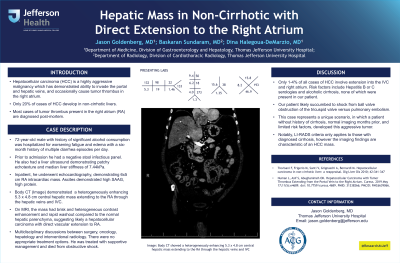
Case Description/Methods: 72-year-old male with significant alcohol consumption was hospitalized for worsening fatigue and edema and had a six-month history of multiple diarrhea episodes per day. He had a negative infectious stool panel and modestly abnormal LFTs (ALP ~300 IU/L, total bilirubin 1.5 mg/dL). His liver demonstrated patchy echotexture with a median liver stiffness value of 7.44 kPa. Due to deteriorating clinical status, including new onset ascites during a routine clinic visit, he was sent for urgent inpatient evaluation. On echocardiography, he had a 6 x 5 cm Right Atrial (RA) intracardiac mass. His ascites had high protein and SAAG levels. Body CT showed a heterogeneously enhancing 5.3 x 4.8 cm central hepatic mass extending to the RA through the hepatic veins and IVC. On MRI, the mass had brisk and heterogeneous contrast enhancement and rapid washout compared to the normal hepatic parenchyma, suggesting likely a hepatocellular carcinoma with direct vascular extension to RA. Multidisciplinary discussions between surgery, oncology, hepatology and interventional radiology. There were no appropriate treatment options. Hence, providers managed with supportive measures in consultation with the patient and family. He decompensated in the following days and died from obstructive shock.
Discussion: Only 1-4% of all cases of HCC involve extension into the IVC and right atrium. Risk factors of this include hepatitis B or C serologies, and alcoholic cirrhosis, none of which our patient carried. Our patient likely succumbed to shock from ball valve obstruction of the tricuspid valve versus pulmonary embolism. This case represents a unique scenario, in which a patient without history of cirrhosis, normal imaging months prior, and limited risk factors, developed this aggressive tumor. LI-RADS criteria only applies to those with diagnosed cirrhosis, however the imaging findings are characteristic of an HCC mass .

Disclosures:
Jason Goldenberg, MD, Baskaran Sundaram, MD, Dina Halegoua-DeMarzio, MD. P1038 - Hepatic Mass in Non-Cirrhotic With Direct Extension to the Right Atrium, ACG 2023 Annual Scientific Meeting Abstracts. Vancouver, BC, Canada: American College of Gastroenterology.
Jason Goldenberg, MD, Baskaran Sundaram, MD, Dina Halegoua-DeMarzio, MD
Thomas Jefferson University Hospital, Philadelphia, PA
Introduction: Hepatocellular carcinoma (HCC) is a highly aggressive malignancy which has demonstrated ability to invade the portal and hepatic veins, and occasionally cause tumor thrombus in the right atrium. Most cases with tumor thrombus present in the right atrium are diagnosed post-mortem. Only 20% of cases of HCC develop in non-cirrhotic livers. We present a case of HCC in a non-cirrhotic patient, initially diagnosed on echocardiogram performed for new onset ascites and lower extremity swelling in the setting of elevated transaminases.
Case Description/Methods: 72-year-old male with significant alcohol consumption was hospitalized for worsening fatigue and edema and had a six-month history of multiple diarrhea episodes per day. He had a negative infectious stool panel and modestly abnormal LFTs (ALP ~300 IU/L, total bilirubin 1.5 mg/dL). His liver demonstrated patchy echotexture with a median liver stiffness value of 7.44 kPa. Due to deteriorating clinical status, including new onset ascites during a routine clinic visit, he was sent for urgent inpatient evaluation. On echocardiography, he had a 6 x 5 cm Right Atrial (RA) intracardiac mass. His ascites had high protein and SAAG levels. Body CT showed a heterogeneously enhancing 5.3 x 4.8 cm central hepatic mass extending to the RA through the hepatic veins and IVC. On MRI, the mass had brisk and heterogeneous contrast enhancement and rapid washout compared to the normal hepatic parenchyma, suggesting likely a hepatocellular carcinoma with direct vascular extension to RA. Multidisciplinary discussions between surgery, oncology, hepatology and interventional radiology. There were no appropriate treatment options. Hence, providers managed with supportive measures in consultation with the patient and family. He decompensated in the following days and died from obstructive shock.
Discussion: Only 1-4% of all cases of HCC involve extension into the IVC and right atrium. Risk factors of this include hepatitis B or C serologies, and alcoholic cirrhosis, none of which our patient carried. Our patient likely succumbed to shock from ball valve obstruction of the tricuspid valve versus pulmonary embolism. This case represents a unique scenario, in which a patient without history of cirrhosis, normal imaging months prior, and limited risk factors, developed this aggressive tumor. LI-RADS criteria only applies to those with diagnosed cirrhosis, however the imaging findings are characteristic of an HCC mass .

Figure: Body CT showing a heterogeneously enhancing 5.3 x 4.8 cm central hepatic mass extending to the RA through the hepatic veins and IVC
Disclosures:
Jason Goldenberg indicated no relevant financial relationships.
Baskaran Sundaram indicated no relevant financial relationships.
Dina Halegoua-DeMarzio: Pfizer – Advisory Committee/Board Member.
Jason Goldenberg, MD, Baskaran Sundaram, MD, Dina Halegoua-DeMarzio, MD. P1038 - Hepatic Mass in Non-Cirrhotic With Direct Extension to the Right Atrium, ACG 2023 Annual Scientific Meeting Abstracts. Vancouver, BC, Canada: American College of Gastroenterology.

