Sunday Poster Session
Category: Liver
P1059 - Hepatocellular Carcinoma With Invasion in Common Bile Duct Presenting With Obstructive Jaundice
Sunday, October 22, 2023
3:30 PM - 7:00 PM PT
Location: Exhibit Hall

Has Audio

Ellen C. Tan, DO
OhioHealth Riverside Methodist Hospital
Columbus, OH
Presenting Author(s)
Ellen C. Tan, DO1, Ruchi Bhatia, MD2, Adam J. Hanje, MD2, Anthony Michaels, MD2, David Y. Lo, MD, FACG2
1OhioHealth Riverside Methodist Hospital, Columbus, OH; 2Ohio Gastroenterology Group, Inc., Columbus, OH
Introduction: Patients with hepatocellular carcinoma (HCC) can present in a variety of ways, from an asymptomatic incidental finding on imaging, to overt liver decompensation. Obstructive jaundice as a first presenting sign of HCC is uncommon.
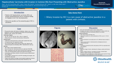
Case Description/Methods: Our case involves a 70-year-old woman with history of diabetes, chronic hepatitis B, tobacco use, and recent gallstone pancreatitis who presented to the hospital with epigastric pain, nausea and vomiting. Labs showed AST 95, ALT 24, ALP 110, DB 0.7, TB 1.1, lipase 67. Ultrasound was concerning for acalculous cholecystitis, leading to cholecystectomy with resultant bile leak. Intraoperatively, her liver appeared nodular. Post-operative MRCP showed diffuse biliary duct dilation with stones, and portal vein thrombus (PVT). AFP was 15160 but contrast imaging was contraindicated initially due to acute kidney injury. EUS showed a left lobe liver mass which underwent fine needle aspiration/biopsy. ERCP demonstrated a long biliary cast with tissue that was extracted and collected (A, B); two 10 French biliary stents were placed given the bile leak. Liver biopsy and biliary contents returned as poorly differentiated HCC (C). Subsequent contrast MRI demonstrated multifocal HCC with PVT extending into the hepatic vein and inferior vena cava. Surgical and medical oncology recommended neoadjuvant immune therapy (atezolizumab and bevacizumab) followed by Y-90.
Discussion: HCC is the fourth leading cause of cancer-related deaths worldwide. Incidence and death rates have increased over the past ten years in the Western countries due to hepatitis C, chronic alcohol use, and metabolic-associated fatty liver disease. In Asia and Africa, hepatitis B remains the most frequent etiology. HCC rarely involves the biliary tree. Obstructive jaundice as a main presentation of HCC occurs in 1-12% of patients. Ductal involvement is not easily seen on CT or MRI and may be noted on ERCP. Due to rarity and subtle appearance, tumors can be missed or misinterpreted as cholangiocarcinoma or choledocholithiasis. Intraductal involvement by HCC and presence of PVT are associated with a poor prognosis. Treatment of HCC is complex and depends on stage of tumor, underlying liver disease, and patient performance status. Options include surgical resection, liver transplantation, locoregional therapies, external beam radiation and systemic therapy. Our patient had contraindications to those options except systemic therapy and radiation, with consideration of Y-90 in the future.

Disclosures:
Ellen C. Tan, DO1, Ruchi Bhatia, MD2, Adam J. Hanje, MD2, Anthony Michaels, MD2, David Y. Lo, MD, FACG2. P1059 - Hepatocellular Carcinoma With Invasion in Common Bile Duct Presenting With Obstructive Jaundice, ACG 2023 Annual Scientific Meeting Abstracts. Vancouver, BC, Canada: American College of Gastroenterology.
1OhioHealth Riverside Methodist Hospital, Columbus, OH; 2Ohio Gastroenterology Group, Inc., Columbus, OH
Introduction: Patients with hepatocellular carcinoma (HCC) can present in a variety of ways, from an asymptomatic incidental finding on imaging, to overt liver decompensation. Obstructive jaundice as a first presenting sign of HCC is uncommon.
Case Description/Methods: Our case involves a 70-year-old woman with history of diabetes, chronic hepatitis B, tobacco use, and recent gallstone pancreatitis who presented to the hospital with epigastric pain, nausea and vomiting. Labs showed AST 95, ALT 24, ALP 110, DB 0.7, TB 1.1, lipase 67. Ultrasound was concerning for acalculous cholecystitis, leading to cholecystectomy with resultant bile leak. Intraoperatively, her liver appeared nodular. Post-operative MRCP showed diffuse biliary duct dilation with stones, and portal vein thrombus (PVT). AFP was 15160 but contrast imaging was contraindicated initially due to acute kidney injury. EUS showed a left lobe liver mass which underwent fine needle aspiration/biopsy. ERCP demonstrated a long biliary cast with tissue that was extracted and collected (A, B); two 10 French biliary stents were placed given the bile leak. Liver biopsy and biliary contents returned as poorly differentiated HCC (C). Subsequent contrast MRI demonstrated multifocal HCC with PVT extending into the hepatic vein and inferior vena cava. Surgical and medical oncology recommended neoadjuvant immune therapy (atezolizumab and bevacizumab) followed by Y-90.
Discussion: HCC is the fourth leading cause of cancer-related deaths worldwide. Incidence and death rates have increased over the past ten years in the Western countries due to hepatitis C, chronic alcohol use, and metabolic-associated fatty liver disease. In Asia and Africa, hepatitis B remains the most frequent etiology. HCC rarely involves the biliary tree. Obstructive jaundice as a main presentation of HCC occurs in 1-12% of patients. Ductal involvement is not easily seen on CT or MRI and may be noted on ERCP. Due to rarity and subtle appearance, tumors can be missed or misinterpreted as cholangiocarcinoma or choledocholithiasis. Intraductal involvement by HCC and presence of PVT are associated with a poor prognosis. Treatment of HCC is complex and depends on stage of tumor, underlying liver disease, and patient performance status. Options include surgical resection, liver transplantation, locoregional therapies, external beam radiation and systemic therapy. Our patient had contraindications to those options except systemic therapy and radiation, with consideration of Y-90 in the future.

Figure: A. Long biliary cast on ERCP B. Extracted biliary cast tissue C. Poorly differentiated HCC on biliary contents
Disclosures:
Ellen Tan indicated no relevant financial relationships.
Ruchi Bhatia indicated no relevant financial relationships.
Adam Hanje: Intercept – Speakers Bureau. Salix – Speakers Bureau.
Anthony Michaels indicated no relevant financial relationships.
David Lo indicated no relevant financial relationships.
Ellen C. Tan, DO1, Ruchi Bhatia, MD2, Adam J. Hanje, MD2, Anthony Michaels, MD2, David Y. Lo, MD, FACG2. P1059 - Hepatocellular Carcinoma With Invasion in Common Bile Duct Presenting With Obstructive Jaundice, ACG 2023 Annual Scientific Meeting Abstracts. Vancouver, BC, Canada: American College of Gastroenterology.
