Sunday Poster Session
Category: Liver
P1080 - Large Microcystic Appearance of Centilobular Venous Congestion in a Case of Acute on Chronic Budd-Chiari Syndrome
Sunday, October 22, 2023
3:30 PM - 7:00 PM PT
Location: Exhibit Hall

Has Audio

Vishwesh Bharadiya
All India Institute of Medical Sciences
Delhi, Delhi, India
Presenting Author(s)
Vishwesh Bharadiya, 1, Umang Arora, MBBS, MD1, Shivanand Gamangatti, MBBS, MD1, Manas Vaishnav, MBBS, MD2, Dr. Shalimar, MBBS, MD2
1All India Institute of Medical Sciences, Delhi, Delhi, India; 2All India Institute of Medical Sciences, New Delhi, Delhi, India
Introduction: Budd-Chiari syndrome (BCS) is characterized by obstruction of the hepatic venous outflow that can lead to hepatic congestion, portal hypertension with ascites, and eventually cirrhosis. While BCS typically presents with well-known features such as ascites and hepatomegaly, atypical presentations can pose diagnostic challenges. Regenerative nodules are often seen on cross-sectional imaging that may be difficult to differentiate from hepatocellular carcinoma. Herein, we present a large microcystic lesion, raising concern for hydatid disease, resulting from acute-on-chronic centrilobular venous congestion due to the progression of venous obstruction.
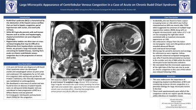
Case Description/Methods: A 22-year-old female was diagnosed with Budd Chiari syndrome when she developed ascites and oesophageal varices six years prior and underwent IVC angioplasty for an IVC web. Pro-coagulant state workup was positive for Protein C deficiency. She clinically improved and was maintained on anticoagulation. She developed a recurrence of ascites and was found to have an absence of patent hepatic veins on ultrasound (USG) doppler and was considered to have progression of BCS to a diffuse disease and was planned for transjugular intrahepatic portosystemic shunt (TIPSS) for the congestion. Incidentally, she was found to have a space-occupying lesion on USG which was further characterized on MRI one month after TIPSS which suggested the presence of multiple large regenerative nodules (RGN) along with an irregular microvesicular cystic lesion of 17 x 10 cm size occupying the right lobe which correlated with an isoechoic microcystic appearance on USG. She underwent a biopsy from the lesion via transjugular route and USG guidance which revealed advanced fibrosis and centrizonal hemorrhage. Microscopy for parasitic elements and serology for echinococcus were negative. Follow-up contrast-enhanced computed tomography scan after 9 months demonstrated an increase in the number and size of RGN while the initial microcystic lesion had become indistinct. We concluded that the microcystic appearance of the right lobe of the liver was due to acute on chronic congestion in this case of BCS which was relieved following TIPSS.
Discussion: This case underscores the importance of recognizing atypical manifestations of BCS and considering acute-on-chronic congestion as a potential etiology for large microcystic lesions in the liver, which can resolve spontaneously upon relief of the congestion.

Disclosures:
Vishwesh Bharadiya, 1, Umang Arora, MBBS, MD1, Shivanand Gamangatti, MBBS, MD1, Manas Vaishnav, MBBS, MD2, Dr. Shalimar, MBBS, MD2. P1080 - Large Microcystic Appearance of Centilobular Venous Congestion in a Case of Acute on Chronic Budd-Chiari Syndrome, ACG 2023 Annual Scientific Meeting Abstracts. Vancouver, BC, Canada: American College of Gastroenterology.
1All India Institute of Medical Sciences, Delhi, Delhi, India; 2All India Institute of Medical Sciences, New Delhi, Delhi, India
Introduction: Budd-Chiari syndrome (BCS) is characterized by obstruction of the hepatic venous outflow that can lead to hepatic congestion, portal hypertension with ascites, and eventually cirrhosis. While BCS typically presents with well-known features such as ascites and hepatomegaly, atypical presentations can pose diagnostic challenges. Regenerative nodules are often seen on cross-sectional imaging that may be difficult to differentiate from hepatocellular carcinoma. Herein, we present a large microcystic lesion, raising concern for hydatid disease, resulting from acute-on-chronic centrilobular venous congestion due to the progression of venous obstruction.
Case Description/Methods: A 22-year-old female was diagnosed with Budd Chiari syndrome when she developed ascites and oesophageal varices six years prior and underwent IVC angioplasty for an IVC web. Pro-coagulant state workup was positive for Protein C deficiency. She clinically improved and was maintained on anticoagulation. She developed a recurrence of ascites and was found to have an absence of patent hepatic veins on ultrasound (USG) doppler and was considered to have progression of BCS to a diffuse disease and was planned for transjugular intrahepatic portosystemic shunt (TIPSS) for the congestion. Incidentally, she was found to have a space-occupying lesion on USG which was further characterized on MRI one month after TIPSS which suggested the presence of multiple large regenerative nodules (RGN) along with an irregular microvesicular cystic lesion of 17 x 10 cm size occupying the right lobe which correlated with an isoechoic microcystic appearance on USG. She underwent a biopsy from the lesion via transjugular route and USG guidance which revealed advanced fibrosis and centrizonal hemorrhage. Microscopy for parasitic elements and serology for echinococcus were negative. Follow-up contrast-enhanced computed tomography scan after 9 months demonstrated an increase in the number and size of RGN while the initial microcystic lesion had become indistinct. We concluded that the microcystic appearance of the right lobe of the liver was due to acute on chronic congestion in this case of BCS which was relieved following TIPSS.
Discussion: This case underscores the importance of recognizing atypical manifestations of BCS and considering acute-on-chronic congestion as a potential etiology for large microcystic lesions in the liver, which can resolve spontaneously upon relief of the congestion.

Figure: MRI abdomen (Axial T1 image, A; Axial T2 weighted image, B; and Coronal T2 weighted image, C) post TIPSS showing a large irregular microvesicular cystic lesion involving the entire right lobe and caudate lobe, appearing T1/T2 isointense with vessels seen coursing within, showing slow progressive enhancement and patchy diffusion restriction.
Disclosures:
Vishwesh Bharadiya indicated no relevant financial relationships.
Umang Arora indicated no relevant financial relationships.
Shivanand Gamangatti indicated no relevant financial relationships.
Manas Vaishnav indicated no relevant financial relationships.
Dr. Shalimar indicated no relevant financial relationships.
Vishwesh Bharadiya, 1, Umang Arora, MBBS, MD1, Shivanand Gamangatti, MBBS, MD1, Manas Vaishnav, MBBS, MD2, Dr. Shalimar, MBBS, MD2. P1080 - Large Microcystic Appearance of Centilobular Venous Congestion in a Case of Acute on Chronic Budd-Chiari Syndrome, ACG 2023 Annual Scientific Meeting Abstracts. Vancouver, BC, Canada: American College of Gastroenterology.
