Sunday Poster Session
Category: Liver
P1104 - Anchoring Bias in the Evaluation of Cholestasis: When Gallstone Disease Delays the Diagnosis of Hepatic Infiltration by Metastatic Melanoma of Unknown Primary
Sunday, October 22, 2023
3:30 PM - 7:00 PM PT
Location: Exhibit Hall

Has Audio

Estefania Flores, MD
University of Iowa Hospitals & Clinics
Iowa City, IA
Presenting Author(s)
Estefania Flores-Cordova, MD, Antonio Sanchez, MD, Ahmed Elkafrawy, MD
University of Iowa Hospitals & Clinics, Iowa City, IA
Introduction: Approximately 3% of melanomas present with distant metastases with no known primary site. Widespread infiltration of the liver is unusual, and its diagnosis can be challenging as imaging and clinical presentation often do not reveal this type of infiltration pattern. We present the case of a patient with abdominal pain and deranged liver enzymes, initially thought to be from gallstone disease.
Case Description/Methods: A 57-year-old man who underwent a cholecystectomy after a 6-week history of right upper quadrant pain, intermittent nausea, and emesis. On presentation, total bilirubin (TB) was 1.4 mg/dL, aspartate transaminase (AST) 61 U/L, aspartate transaminase (ALT) 133 U/L, alkaline phosphatase (ALP) 424 U/L. Ultrasound showed multiple gallstones and marked heterogeneity of the liver parenchyma. During laparoscopic cholecystectomy, he was noted to have a “cystic” liver. The procedure was completed uneventfully.
Ten days postoperatively, he presented to our academic center with nausea, vomiting, and abdominal pain. Abdominal exam revealed right upper quadrant tenderness. TB 4.0 mg/dL, ALT 68 U/L, AST 56 U/L, ALP 450 U/L, INR 1.3. Computerized tomography showed lobulated hepatic contour, and perihepatic fluid collection concerning for a biloma. Endoscopic retrograde cholangiopancreatography (ERCP) was performed given the concern for a biliary leak, but it was unremarkable. An abdominal MRI showed moderate hepatomegaly and diffuse hepatic parenchymal nodularity. Percutaneous US-guided liver biopsy was performed, positive for infiltrative metastatic melanoma. A complete dermatologic and ocular exam did not show any concerning lesions. The patient was started on encorafenib and binimetinib. His clinical status rapidly deteriorated TB increased up to 11 mg/dL and the patient died 3 days later.
Discussion: Malignant melanoma may involve any organ and while it commonly metastasizes to the lymph nodes, lungs, liver, brain, or bone, diffuse hepatic infiltration is rare but fatal. The diagnosis of widespread liver metastasis with unknown primary is challenging as standard laboratory values are not helpful in identifying the presence of malignancy. When the etiology of cholestatic liver injury is unclear, diffuse metastatic tumor infiltration from metastatic melanoma of unknown primary should be considered in the differential, early liver biopsy is essential in establishing the diagnosis and prognosis.

Disclosures:
Estefania Flores-Cordova, MD, Antonio Sanchez, MD, Ahmed Elkafrawy, MD. P1104 - Anchoring Bias in the Evaluation of Cholestasis: When Gallstone Disease Delays the Diagnosis of Hepatic Infiltration by Metastatic Melanoma of Unknown Primary, ACG 2023 Annual Scientific Meeting Abstracts. Vancouver, BC, Canada: American College of Gastroenterology.
University of Iowa Hospitals & Clinics, Iowa City, IA
Introduction: Approximately 3% of melanomas present with distant metastases with no known primary site. Widespread infiltration of the liver is unusual, and its diagnosis can be challenging as imaging and clinical presentation often do not reveal this type of infiltration pattern. We present the case of a patient with abdominal pain and deranged liver enzymes, initially thought to be from gallstone disease.
Case Description/Methods: A 57-year-old man who underwent a cholecystectomy after a 6-week history of right upper quadrant pain, intermittent nausea, and emesis. On presentation, total bilirubin (TB) was 1.4 mg/dL, aspartate transaminase (AST) 61 U/L, aspartate transaminase (ALT) 133 U/L, alkaline phosphatase (ALP) 424 U/L. Ultrasound showed multiple gallstones and marked heterogeneity of the liver parenchyma. During laparoscopic cholecystectomy, he was noted to have a “cystic” liver. The procedure was completed uneventfully.
Ten days postoperatively, he presented to our academic center with nausea, vomiting, and abdominal pain. Abdominal exam revealed right upper quadrant tenderness. TB 4.0 mg/dL, ALT 68 U/L, AST 56 U/L, ALP 450 U/L, INR 1.3. Computerized tomography showed lobulated hepatic contour, and perihepatic fluid collection concerning for a biloma. Endoscopic retrograde cholangiopancreatography (ERCP) was performed given the concern for a biliary leak, but it was unremarkable. An abdominal MRI showed moderate hepatomegaly and diffuse hepatic parenchymal nodularity. Percutaneous US-guided liver biopsy was performed, positive for infiltrative metastatic melanoma. A complete dermatologic and ocular exam did not show any concerning lesions. The patient was started on encorafenib and binimetinib. His clinical status rapidly deteriorated TB increased up to 11 mg/dL and the patient died 3 days later.
Discussion: Malignant melanoma may involve any organ and while it commonly metastasizes to the lymph nodes, lungs, liver, brain, or bone, diffuse hepatic infiltration is rare but fatal. The diagnosis of widespread liver metastasis with unknown primary is challenging as standard laboratory values are not helpful in identifying the presence of malignancy. When the etiology of cholestatic liver injury is unclear, diffuse metastatic tumor infiltration from metastatic melanoma of unknown primary should be considered in the differential, early liver biopsy is essential in establishing the diagnosis and prognosis.

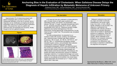
Figure: A: Diffuse hepatic parenchymal nodularity on MRI.
B. PET -CT with hepatomegaly with numerous radiotracer-avid lesions.
C and D. Liver biopsy specimen: The tumor cells show brown-pigment cytoplasmic pigmentation, which is consistent with malignant melanoma (A, hematoxylin and eosin ×100)
B. PET -CT with hepatomegaly with numerous radiotracer-avid lesions.
C and D. Liver biopsy specimen: The tumor cells show brown-pigment cytoplasmic pigmentation, which is consistent with malignant melanoma (A, hematoxylin and eosin ×100)
Disclosures:
Estefania Flores-Cordova indicated no relevant financial relationships.
Antonio Sanchez: Abbvie – Grant/Research Support. Arrowhead – Grant/Research Support. Boehringer-Ingelheim – Grant/Research Support. Escient – Grant/Research Support. Gilead – Grant/Research Support. Intercept Pharmaceuticals – Grant/Research Support. Inventiva – Grant/Research Support. Merck – Grant/Research Support.
Ahmed Elkafrawy indicated no relevant financial relationships.
Estefania Flores-Cordova, MD, Antonio Sanchez, MD, Ahmed Elkafrawy, MD. P1104 - Anchoring Bias in the Evaluation of Cholestasis: When Gallstone Disease Delays the Diagnosis of Hepatic Infiltration by Metastatic Melanoma of Unknown Primary, ACG 2023 Annual Scientific Meeting Abstracts. Vancouver, BC, Canada: American College of Gastroenterology.
