Sunday Poster Session
Category: Liver
P1119 - Evading Death – Recurrent Ascending Cholangitis Secondary to Multiple Ductal Plate Malformations
Sunday, October 22, 2023
3:30 PM - 7:00 PM PT
Location: Exhibit Hall

Has Audio
.jpg)
Adwait Patel, MD
Bayonne Medical Center
BAYONNE, NJ
Presenting Author(s)
Adwait Patel, MD1, Neil Sondhi, MD2, Dhanush Hoskere, DO3, Jennifer Hsieh, MD4, John Dedousis, MD5, Antonio Tsompanidis, DO5
1Bayonne Medical Center, Harrison, NJ; 2Hoboken University Medical Centerr, Marlboro, NJ; 3CarePoint Health, Bayonne, NJ; 4Carepoint Medical Group, Hoboken, NJ; 5Bayonne Medical Center, Bayonne, NJ
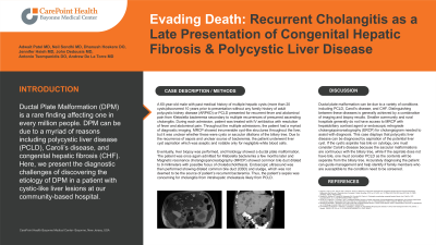
Introduction: Ductal Plate Malformation (DPM) is a rare finding affecting one in every million people. DPM can be due to a myriad of reasons including polycystic liver disease (PCLD), Caroli’s disease, and congenital hepatic fibrosis (CHF). Here, we present the diagnostic challenges of discovering the etiology of DPM in a patient with cystic-like liver lesions at our community-based hospital.
Case Description/Methods: 60-year-old male with past medical history of multiple hepatic cysts discovered 10 years ago without any family history of adult polycystic kidney disease (APCKD) or PCLD presented for recurrent fever and abdominal pain from Klebsiella pneumonia bacteremia secondary to multiple recurrences of presumed ascending cholangitis. Throughout the multiple admissions, each treated with intravenous antibiotics, the patient had a myriad of diagnostic imaging. Magnetic resonance cholangiopancreatography (MRCP) with gadolinium showed innumerable cyst-like lesions throughout the liver, but it was unclear whether these were cysts or saccular dilations of the biliary tree. Due to unclear source of bacteremia, patient underwent liver cyst aspiration which was aseptic and notable only for negligible white blood cells. Eventually, liver biopsy was performed, and histology showed ductal plate malformation. Endoscopic ultrasound was then performed showing dilated CBD and sludge, which was not deemed to be the source of patient’s recurrent bacteremia. Thus, the patient’s sepsis was concerning for cholangitis from intrahepatic cholestasis likely from PCLD.
Discussion: Ductal plate malformation can be due to a variety of conditions including PCLD, Caroli’s disease, and CHF. Distinguishing between these diseases is generally achieved by a combination of imaging and biopsy results. Smaller community and rural hospitals generally do not have access to MRCP with hepatobiliary contrast agent or endoscopic retrograde cholangiopancreatography (ERCP) for cholangiogram needed to assist with diagnosis. This case displays that polycystic liver disease can be diagnosed by aspiration of the potential liver cyst. If the cystic aspirate has bile on cytology, one must consider Caroli’s disease because the saccular malformations are continuous with the biliary tree, while if the aspirate does not have bile, one must consider PCLD as the contents will be separate from the biliary tree. Accurately diagnosing the patient can guide management and help identify if family members who are susceptible to the condition need to be screened.

Disclosures:
Adwait Patel, MD1, Neil Sondhi, MD2, Dhanush Hoskere, DO3, Jennifer Hsieh, MD4, John Dedousis, MD5, Antonio Tsompanidis, DO5. P1119 - Evading Death – Recurrent Ascending Cholangitis Secondary to Multiple Ductal Plate Malformations, ACG 2023 Annual Scientific Meeting Abstracts. Vancouver, BC, Canada: American College of Gastroenterology.
1Bayonne Medical Center, Harrison, NJ; 2Hoboken University Medical Centerr, Marlboro, NJ; 3CarePoint Health, Bayonne, NJ; 4Carepoint Medical Group, Hoboken, NJ; 5Bayonne Medical Center, Bayonne, NJ
Introduction: Ductal Plate Malformation (DPM) is a rare finding affecting one in every million people. DPM can be due to a myriad of reasons including polycystic liver disease (PCLD), Caroli’s disease, and congenital hepatic fibrosis (CHF). Here, we present the diagnostic challenges of discovering the etiology of DPM in a patient with cystic-like liver lesions at our community-based hospital.
Case Description/Methods: 60-year-old male with past medical history of multiple hepatic cysts discovered 10 years ago without any family history of adult polycystic kidney disease (APCKD) or PCLD presented for recurrent fever and abdominal pain from Klebsiella pneumonia bacteremia secondary to multiple recurrences of presumed ascending cholangitis. Throughout the multiple admissions, each treated with intravenous antibiotics, the patient had a myriad of diagnostic imaging. Magnetic resonance cholangiopancreatography (MRCP) with gadolinium showed innumerable cyst-like lesions throughout the liver, but it was unclear whether these were cysts or saccular dilations of the biliary tree. Due to unclear source of bacteremia, patient underwent liver cyst aspiration which was aseptic and notable only for negligible white blood cells. Eventually, liver biopsy was performed, and histology showed ductal plate malformation. Endoscopic ultrasound was then performed showing dilated CBD and sludge, which was not deemed to be the source of patient’s recurrent bacteremia. Thus, the patient’s sepsis was concerning for cholangitis from intrahepatic cholestasis likely from PCLD.
Discussion: Ductal plate malformation can be due to a variety of conditions including PCLD, Caroli’s disease, and CHF. Distinguishing between these diseases is generally achieved by a combination of imaging and biopsy results. Smaller community and rural hospitals generally do not have access to MRCP with hepatobiliary contrast agent or endoscopic retrograde cholangiopancreatography (ERCP) for cholangiogram needed to assist with diagnosis. This case displays that polycystic liver disease can be diagnosed by aspiration of the potential liver cyst. If the cystic aspirate has bile on cytology, one must consider Caroli’s disease because the saccular malformations are continuous with the biliary tree, while if the aspirate does not have bile, one must consider PCLD as the contents will be separate from the biliary tree. Accurately diagnosing the patient can guide management and help identify if family members who are susceptible to the condition need to be screened.

Figure: Innumerable cysts scattered through out the liver
Disclosures:
Adwait Patel indicated no relevant financial relationships.
Neil Sondhi indicated no relevant financial relationships.
Dhanush Hoskere indicated no relevant financial relationships.
Jennifer Hsieh indicated no relevant financial relationships.
John Dedousis indicated no relevant financial relationships.
Antonio Tsompanidis indicated no relevant financial relationships.
Adwait Patel, MD1, Neil Sondhi, MD2, Dhanush Hoskere, DO3, Jennifer Hsieh, MD4, John Dedousis, MD5, Antonio Tsompanidis, DO5. P1119 - Evading Death – Recurrent Ascending Cholangitis Secondary to Multiple Ductal Plate Malformations, ACG 2023 Annual Scientific Meeting Abstracts. Vancouver, BC, Canada: American College of Gastroenterology.
