Sunday Poster Session
Category: Liver
P1120 - It’s Raining Clots: Hemobilia a Rare Complication of Hepatocellular Carcinoma
Sunday, October 22, 2023
3:30 PM - 7:00 PM PT
Location: Exhibit Hall

Has Audio
.jpg)
Adwait Patel, MD
Bayonne Medical Center
BAYONNE, NJ
Presenting Author(s)
Adwait Patel, MD1, Neil Sondhi, MD2, Dhanush Hoskere, DO3, Jennifer Hsieh, MD4, John Dedousis, MD5, Antonio Tsompanidis, DO5
1Bayonne Medical Center, Harrison, NJ; 2Hoboken University Medical Centerr, Marlboro, NJ; 3CarePoint Health, Bayonne, NJ; 4Carepoint Medical Group, Hoboken, NJ; 5Bayonne Medical Center, Bayonne, NJ
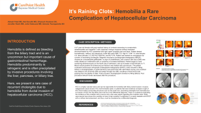
Introduction: Hemobilia is defined as bleeding from the biliary tract and is an uncommon but important cause of gastrointestinal hemorrhage. Hemobilia predominantly is iatrogenic and is often precipitated by invasive procedures involving the liver, pancreas, or biliary tree. Here, we present a rare case of recurrent cholangitis due to hemobilia from ductal invasion of hepatocellular carcinoma (HCC).
Case Description/Methods: A 47-year-old female with past medical history of cirrhosis secondary to nonalcoholic steatohepatitis and hepatitis C with sustained virologic response without treatment decompensated by HCC presented with right upper quadrant pain and fever. Patient denied hematemesis, melena, and hematuria. Initial labs were WBC 2.5, Hgb 9.7, INR 1.4, total bilirubin 4.9, direct bilirubin 3.3, AST/ALT 608/184, and ALP 376. Patient was started on meropenem for concern of ascending cholangitis. Magnetic resonance cholangiopancreatography (MRCP) showed an unremarkable gallbladder, no sign of cholelithiasis, and common bile duct (CBD) was mildly dilated at 9 millimeters with no evidence of choledocholithiasis. Patient began to have worsening abdominal pain on the fourth day of admission, and an increase in total bilirubin to 11. Blood culture positive for Enterococcus Faecium was treated with vancomycin. The patient underwent endoscopic retrograde cholangiopancreatography (ERCP), which showed a filling defect in cholangiography. A sphincterotomy was performed with self-limited oozing, and a balloon was dilated in the proximal CBD and swept through the CBD, resulting in frank blood clots passing from the papilla of Vater. Final occlusion cholangiogram showed no filling defects. Post procedure bilirubin levels decreased after the procedure.
Discussion: HCC is a highly vascular tumor and can be responsible for hemobilia with biliary ductal invasion. Invasive malignancies cause around 10% of all hemobilia cases. In patients that have evidence of sepsis in light of HCC that invades surrounding structures such as the portal vein, ascending cholangitis from hemobilia due to invasion of HCC into the biliary tree must be on the differential diagnosis. There is no literature estimating the incidence of this condition and is rare as only a few case reports regarding this condition exist. While case reports have shown this condition to have a high mortality if undetected, it is easily treatable with ERCP with or without stent placement, and thus, must be considered as a differential diagnosis.

Disclosures:
Adwait Patel, MD1, Neil Sondhi, MD2, Dhanush Hoskere, DO3, Jennifer Hsieh, MD4, John Dedousis, MD5, Antonio Tsompanidis, DO5. P1120 - It’s Raining Clots: Hemobilia a Rare Complication of Hepatocellular Carcinoma, ACG 2023 Annual Scientific Meeting Abstracts. Vancouver, BC, Canada: American College of Gastroenterology.
1Bayonne Medical Center, Harrison, NJ; 2Hoboken University Medical Centerr, Marlboro, NJ; 3CarePoint Health, Bayonne, NJ; 4Carepoint Medical Group, Hoboken, NJ; 5Bayonne Medical Center, Bayonne, NJ
Introduction: Hemobilia is defined as bleeding from the biliary tract and is an uncommon but important cause of gastrointestinal hemorrhage. Hemobilia predominantly is iatrogenic and is often precipitated by invasive procedures involving the liver, pancreas, or biliary tree. Here, we present a rare case of recurrent cholangitis due to hemobilia from ductal invasion of hepatocellular carcinoma (HCC).
Case Description/Methods: A 47-year-old female with past medical history of cirrhosis secondary to nonalcoholic steatohepatitis and hepatitis C with sustained virologic response without treatment decompensated by HCC presented with right upper quadrant pain and fever. Patient denied hematemesis, melena, and hematuria. Initial labs were WBC 2.5, Hgb 9.7, INR 1.4, total bilirubin 4.9, direct bilirubin 3.3, AST/ALT 608/184, and ALP 376. Patient was started on meropenem for concern of ascending cholangitis. Magnetic resonance cholangiopancreatography (MRCP) showed an unremarkable gallbladder, no sign of cholelithiasis, and common bile duct (CBD) was mildly dilated at 9 millimeters with no evidence of choledocholithiasis. Patient began to have worsening abdominal pain on the fourth day of admission, and an increase in total bilirubin to 11. Blood culture positive for Enterococcus Faecium was treated with vancomycin. The patient underwent endoscopic retrograde cholangiopancreatography (ERCP), which showed a filling defect in cholangiography. A sphincterotomy was performed with self-limited oozing, and a balloon was dilated in the proximal CBD and swept through the CBD, resulting in frank blood clots passing from the papilla of Vater. Final occlusion cholangiogram showed no filling defects. Post procedure bilirubin levels decreased after the procedure.
Discussion: HCC is a highly vascular tumor and can be responsible for hemobilia with biliary ductal invasion. Invasive malignancies cause around 10% of all hemobilia cases. In patients that have evidence of sepsis in light of HCC that invades surrounding structures such as the portal vein, ascending cholangitis from hemobilia due to invasion of HCC into the biliary tree must be on the differential diagnosis. There is no literature estimating the incidence of this condition and is rare as only a few case reports regarding this condition exist. While case reports have shown this condition to have a high mortality if undetected, it is easily treatable with ERCP with or without stent placement, and thus, must be considered as a differential diagnosis.

Figure: A) Filling defect seen on cholangiogram in upper third of common bile duct
B) biliary sphincterotomy and balloon sweep with removal of multiple blood clots
B) biliary sphincterotomy and balloon sweep with removal of multiple blood clots
Disclosures:
Adwait Patel indicated no relevant financial relationships.
Neil Sondhi indicated no relevant financial relationships.
Dhanush Hoskere indicated no relevant financial relationships.
Jennifer Hsieh indicated no relevant financial relationships.
John Dedousis indicated no relevant financial relationships.
Antonio Tsompanidis indicated no relevant financial relationships.
Adwait Patel, MD1, Neil Sondhi, MD2, Dhanush Hoskere, DO3, Jennifer Hsieh, MD4, John Dedousis, MD5, Antonio Tsompanidis, DO5. P1120 - It’s Raining Clots: Hemobilia a Rare Complication of Hepatocellular Carcinoma, ACG 2023 Annual Scientific Meeting Abstracts. Vancouver, BC, Canada: American College of Gastroenterology.
