Sunday Poster Session
Category: Small Intestine
P1261 - Cytomegalovirus Duodenitis Causing Partial Small Bowel Obstruction in the Setting of Diffuse Large B-Cell Lymphoma
Sunday, October 22, 2023
3:30 PM - 7:00 PM PT
Location: Exhibit Hall

Has Audio
- MK
Majd Khadra, MD
Detroit Medical Center/Wayne State University
Detroit, MI
Presenting Author(s)
Majd Khadra, MD1, Anshu Wadehra, MD2, Bashar Mohamad, MD2
1Detroit Medical Center/Wayne State University, Detroit, MI; 2Wayne State University, Detroit, MI
Introduction: Duodenitis, the inflammation of the first part of the small intestine, is commonly found in 10% of patients undergoing endoscopy. Cytomegalovirus infection is known to be opportunistic in immunocompromised patients with high morbidity and mortality.
Case Description/Methods: A 48-year-old male with a past medical history of diffuse large B-cell lymphoma diagnosed in 2011 status post CAR-T cell treatment 4 months prior to admission with acute cholecystitis. Patient was admitted to the emergency department with left mandibular swelling and worsening acute right upper quadrant pain associated with nausea. Vital signs on admission were within normal limits. On physical exam, left mandibular swelling and right upper quadrant tenderness was noted. Labs on admission significant for platelet count of 65 K/CUMM and WBC of 1.1 K/CUMM. Chart review revealed patient had cytomegalovirus quantitative PCR of 1068 IU/mL, 5 weeks prior to admission in outpatient setting.
Esophagogastroduodenoscopy revealed benign duodenal stricture noted in the second part of duodenum with edematous submucosa near obstruction of the lumen.
Our patient's cytomegalovirus quantitative PCR increased from 861 IU/ml to 9467 IU/ml in 2 weeks. Esophagogastroduodenoscopy biopsy results of cytomegalovirus duodenitis were revealed concurrently with the cytomegalovirus quantitative PCR level of 9467 IU/ml . IV Ganciclovir was started on the same day. Clinically, the patient improved over the subsequent week with resolved nausea and vomiting. Nasogastric tube was removed and diet was advanced as tolerated. Objectively, cytomegalovirus quantitative PCR levels decreased to less than 137 IU/ml one week after initiating IV Ganciclovir therapy. Based on our case, there could be an association between cytomegalovirus quantitative PCR levels and the degree of cytomegalovirus duodenitis.
Discussion: The diagnostic gold standard for cytomegalovirus infection is histopathological examination of endoscopic biopsy or surgical specimen.
Cytomegalovirus duodenitis should be on the differential diagnosis of immunocompromised patients that develop small bowel obstruction. Diagnosis relies on histopathologic examination of endoscopic biopsy specimens. In our case, there was an improvement in both observed symptoms of small bowel obstruction and cytomegalovirus quantitative PCR level with intravenous Ganciclovir therapy, which may reflect a possible association between levels of viral load and activity of cytomegalovirus duodenitis.

Disclosures:
Majd Khadra, MD1, Anshu Wadehra, MD2, Bashar Mohamad, MD2. P1261 - Cytomegalovirus Duodenitis Causing Partial Small Bowel Obstruction in the Setting of Diffuse Large B-Cell Lymphoma, ACG 2023 Annual Scientific Meeting Abstracts. Vancouver, BC, Canada: American College of Gastroenterology.
1Detroit Medical Center/Wayne State University, Detroit, MI; 2Wayne State University, Detroit, MI
Introduction: Duodenitis, the inflammation of the first part of the small intestine, is commonly found in 10% of patients undergoing endoscopy. Cytomegalovirus infection is known to be opportunistic in immunocompromised patients with high morbidity and mortality.
Case Description/Methods: A 48-year-old male with a past medical history of diffuse large B-cell lymphoma diagnosed in 2011 status post CAR-T cell treatment 4 months prior to admission with acute cholecystitis. Patient was admitted to the emergency department with left mandibular swelling and worsening acute right upper quadrant pain associated with nausea. Vital signs on admission were within normal limits. On physical exam, left mandibular swelling and right upper quadrant tenderness was noted. Labs on admission significant for platelet count of 65 K/CUMM and WBC of 1.1 K/CUMM. Chart review revealed patient had cytomegalovirus quantitative PCR of 1068 IU/mL, 5 weeks prior to admission in outpatient setting.
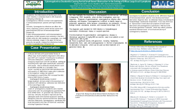
Esophagogastroduodenoscopy revealed benign duodenal stricture noted in the second part of duodenum with edematous submucosa near obstruction of the lumen.
Our patient's cytomegalovirus quantitative PCR increased from 861 IU/ml to 9467 IU/ml in 2 weeks. Esophagogastroduodenoscopy biopsy results of cytomegalovirus duodenitis were revealed concurrently with the cytomegalovirus quantitative PCR level of 9467 IU/ml . IV Ganciclovir was started on the same day. Clinically, the patient improved over the subsequent week with resolved nausea and vomiting. Nasogastric tube was removed and diet was advanced as tolerated. Objectively, cytomegalovirus quantitative PCR levels decreased to less than 137 IU/ml one week after initiating IV Ganciclovir therapy. Based on our case, there could be an association between cytomegalovirus quantitative PCR levels and the degree of cytomegalovirus duodenitis.
Discussion: The diagnostic gold standard for cytomegalovirus infection is histopathological examination of endoscopic biopsy or surgical specimen.
Cytomegalovirus duodenitis should be on the differential diagnosis of immunocompromised patients that develop small bowel obstruction. Diagnosis relies on histopathologic examination of endoscopic biopsy specimens. In our case, there was an improvement in both observed symptoms of small bowel obstruction and cytomegalovirus quantitative PCR level with intravenous Ganciclovir therapy, which may reflect a possible association between levels of viral load and activity of cytomegalovirus duodenitis.

Figure: Benign Duodenal Stricture Noted in the Second Part of Duodenum With Edematous Submucosa Near Obstruction of the Lumen
Disclosures:
Majd Khadra indicated no relevant financial relationships.
Anshu Wadehra indicated no relevant financial relationships.
Bashar Mohamad indicated no relevant financial relationships.
Majd Khadra, MD1, Anshu Wadehra, MD2, Bashar Mohamad, MD2. P1261 - Cytomegalovirus Duodenitis Causing Partial Small Bowel Obstruction in the Setting of Diffuse Large B-Cell Lymphoma, ACG 2023 Annual Scientific Meeting Abstracts. Vancouver, BC, Canada: American College of Gastroenterology.
