Sunday Poster Session
Category: Small Intestine
P1267 - Intermittent Small Bowel Obstruction Due to a Rare Windsock Duodenal Diverticulum
Sunday, October 22, 2023
3:30 PM - 7:00 PM PT
Location: Exhibit Hall

Has Audio

Hassan Ali Al Saleh, DO, MSc
University at Buffalo
Buffalo, NY
Presenting Author(s)
Hassan Ali Al Saleh, DO, MSc, Elizabeth Z. Gregory, MD, Ramon E.. Rivera, MD
University at Buffalo, Buffalo, NY
Introduction: A windsock-type duodenal diverticulum is a rare subtype of intraluminal duodenal diverticula (IDD) that arises due to the incomplete recanalization of the foregut during embryogenesis. Most patients are asymptomatic and non-specific symptoms may present as abdominal distention and early satiety, while more serious complications include bleeding, bowel obstruction, biliary or pancreatic duct manifestations, and rarely perforation. We present a case of a windsock duodenal diverticulum causing significant gastric distention and bowel obstruction that was treated conservatively.
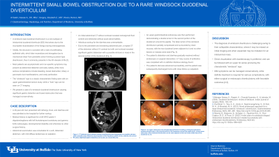
Case Description/Methods: A 48-year-old man presented with lethargy, fever, and diarrhea and was admitted to the hospital for further workup. Medical history is significant for a left WHO grade II oligodendroglioma with left frontotemporal craniotomy and gamma knife radiosurgery, developmental disability and otherwise no other surgical history. Abdominal examination was remarkable for a soft, distended abdomen, with mild diffuse tenderness on palpation. An abdominal CT without contrast revealed rectosigmoid fluid content. Infectious workup for the diarrhea was unremarkable, and due to persistent and worsening abdominal pain, a repeat CT of the abdomen without contrast revealed significant gastric distension with a possible stricture or mass in the second portion of the duodenum [Fig. 1A,B]. An upper gastrointestinal endoscopy was then performed demonstrating a double lumen in the second portion of the duodenum around the papilla [Fig. 1C,D]. The false lumen of the windsock diverticulum partially compressed and surrounded by clean mucosa, with the true duodenal lumen adjacent to it and no other lesions or masses were seen [Fig. 1C,D]. The patient’s distention and diarrhea resolved without endoscopic or surgical intervention. A 7-day course of antibiotics was completed with no definite infectious etiology found, and the patient was subsequently discharged home with close follow up outpatient.
Discussion: The diagnosis of windsock diverticula is challenging owing to their collapsible characteristics, where it may be missed on initial imaging and when expanded may be mistaken for an intraluminal mass. Direct visualization with duodenoscopy is preferred, and can be followed with an upper GI series producing the characteristic “windsock” sign. Mild symptoms can be managed conservatively, while definite treatment is required for serious complications, with either surgical or endoscopic diverticulotomy with favorable outcomes.

Disclosures:
Hassan Ali Al Saleh, DO, MSc, Elizabeth Z. Gregory, MD, Ramon E.. Rivera, MD. P1267 - Intermittent Small Bowel Obstruction Due to a Rare Windsock Duodenal Diverticulum, ACG 2023 Annual Scientific Meeting Abstracts. Vancouver, BC, Canada: American College of Gastroenterology.
University at Buffalo, Buffalo, NY
Introduction: A windsock-type duodenal diverticulum is a rare subtype of intraluminal duodenal diverticula (IDD) that arises due to the incomplete recanalization of the foregut during embryogenesis. Most patients are asymptomatic and non-specific symptoms may present as abdominal distention and early satiety, while more serious complications include bleeding, bowel obstruction, biliary or pancreatic duct manifestations, and rarely perforation. We present a case of a windsock duodenal diverticulum causing significant gastric distention and bowel obstruction that was treated conservatively.
Case Description/Methods: A 48-year-old man presented with lethargy, fever, and diarrhea and was admitted to the hospital for further workup. Medical history is significant for a left WHO grade II oligodendroglioma with left frontotemporal craniotomy and gamma knife radiosurgery, developmental disability and otherwise no other surgical history. Abdominal examination was remarkable for a soft, distended abdomen, with mild diffuse tenderness on palpation. An abdominal CT without contrast revealed rectosigmoid fluid content. Infectious workup for the diarrhea was unremarkable, and due to persistent and worsening abdominal pain, a repeat CT of the abdomen without contrast revealed significant gastric distension with a possible stricture or mass in the second portion of the duodenum [Fig. 1A,B]. An upper gastrointestinal endoscopy was then performed demonstrating a double lumen in the second portion of the duodenum around the papilla [Fig. 1C,D]. The false lumen of the windsock diverticulum partially compressed and surrounded by clean mucosa, with the true duodenal lumen adjacent to it and no other lesions or masses were seen [Fig. 1C,D]. The patient’s distention and diarrhea resolved without endoscopic or surgical intervention. A 7-day course of antibiotics was completed with no definite infectious etiology found, and the patient was subsequently discharged home with close follow up outpatient.
Discussion: The diagnosis of windsock diverticula is challenging owing to their collapsible characteristics, where it may be missed on initial imaging and when expanded may be mistaken for an intraluminal mass. Direct visualization with duodenoscopy is preferred, and can be followed with an upper GI series producing the characteristic “windsock” sign. Mild symptoms can be managed conservatively, while definite treatment is required for serious complications, with either surgical or endoscopic diverticulotomy with favorable outcomes.

Figure: Figure 1. CT abdomen and pelvis without contrast revealing a possible stricture or mass within the second portion of the duodenum (white arrow) (A, B), with signs of gastric distention (B); axial (A) and coronal (B) views. An upper endoscopy was subsequently performed revealing a windsock intraluminal duodenal diverticulum in the second portion of the duodenum around the papilla; a false lumen (black star) of the windsock diverticulum can be seen adjacent to the true lumen (white star) of the duodenum (C, D).
Disclosures:
Hassan Ali Al Saleh indicated no relevant financial relationships.
Elizabeth Gregory indicated no relevant financial relationships.
Ramon Rivera indicated no relevant financial relationships.
Hassan Ali Al Saleh, DO, MSc, Elizabeth Z. Gregory, MD, Ramon E.. Rivera, MD. P1267 - Intermittent Small Bowel Obstruction Due to a Rare Windsock Duodenal Diverticulum, ACG 2023 Annual Scientific Meeting Abstracts. Vancouver, BC, Canada: American College of Gastroenterology.
