Sunday Poster Session
Category: Small Intestine
P1276 - A Case of Complicated Case of Mesenteric Vein Thrombosis
Sunday, October 22, 2023
3:30 PM - 7:00 PM PT
Location: Exhibit Hall

Has Audio

Jonathan Hansen, DO
Kirk Kerkorian School of Medicine at UNLV
Las Vegas, NV
Presenting Author(s)
Jonathan Hansen, DO, Katerina Roma, DO, Lubaba Haque, DO, Kyaw Min Tun, DO, Amith Subhash, MD
Kirk Kerkorian School of Medicine at UNLV, Las Vegas, NV
Introduction: Mesenteric vein thrombosis (MVT) is a rare cause of small bowel ischemia, composing less than 10% of cases. When diagnosed early and treated, mortality is less than 10%. However, in cases where treatment is delayed by 24 hours or more, mortality can be as high as 80%. Herein, we present an unusual case of MVT due to a hypercoagulable state induced by malignancy.
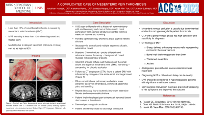
Case Description/Methods: A 65-year-old female with a history of hemicolectomy with end ileostomy and mucus fistula due to cecal perforation from sigmoid stricture presented with two weeks of nausea and vomiting. Flexible sigmoidoscopy showed a distal sigmoid fibrotic stricture that was biopsied. Ileoscopy via stoma found multiple, distinct segments of pale, edematous bowel that were biopsied. Colon stricture biopsy showed poorly differentiated adenocarcinoma while ileoscopy biopsies showed benign small bowel mucosa with superficial erosions. Initial CT showed diffuse wall thickening of the small bowel and superior mesenteric vein (SMV) narrowing concerning for chronic occlusion. However, follow-up CT angiogram (CTA) found a patent SMV with inflammatory changes of the entire small and large bowel (Figure 1). Her hospital course was complicated by pulmonary embolism, lower extremity deep vein thrombosis, continued abdominal pain, and vomiting. Repeat ileoscopy found ischemic ileum with extensive fibrosis and untraversable stricture. It was concluded that she had likely developed pan-ischemia of her small bowel due to venous thrombosis. She was determined to be a poor surgical candidate given the extensive nature of the ischemia. Following extensive family discussion, the patient was ultimately discharged to hospice.
Discussion: Mesenteric venous occlusion is usually due to mechanical obstruction or thrombosis in a hypercoagulable patient. CTA with a portal venous phase has high sensitivity and specificity for diagnosis. CT findings of MVT include sharp, defined enhancing venous walls representing contrast in the vasa vasorum, bowel wall thickening greater than 3mm, thickened mesentery, and ascites. Unfortunately, by the time of diagnosis, our patient’s pan-ischemia was so extensive it was deemed inoperable. This case highlights the difficulty of diagnosing MVT as well as the need to consider MVT in a hypercoagulable patient with significant small bowel edema with no other identifiable causes. Early surgical intervention may have prevented worsening of her symptoms and improved her outcome.

Disclosures:
Jonathan Hansen, DO, Katerina Roma, DO, Lubaba Haque, DO, Kyaw Min Tun, DO, Amith Subhash, MD. P1276 - A Case of Complicated Case of Mesenteric Vein Thrombosis, ACG 2023 Annual Scientific Meeting Abstracts. Vancouver, BC, Canada: American College of Gastroenterology.
Kirk Kerkorian School of Medicine at UNLV, Las Vegas, NV
Introduction: Mesenteric vein thrombosis (MVT) is a rare cause of small bowel ischemia, composing less than 10% of cases. When diagnosed early and treated, mortality is less than 10%. However, in cases where treatment is delayed by 24 hours or more, mortality can be as high as 80%. Herein, we present an unusual case of MVT due to a hypercoagulable state induced by malignancy.
Case Description/Methods: A 65-year-old female with a history of hemicolectomy with end ileostomy and mucus fistula due to cecal perforation from sigmoid stricture presented with two weeks of nausea and vomiting. Flexible sigmoidoscopy showed a distal sigmoid fibrotic stricture that was biopsied. Ileoscopy via stoma found multiple, distinct segments of pale, edematous bowel that were biopsied. Colon stricture biopsy showed poorly differentiated adenocarcinoma while ileoscopy biopsies showed benign small bowel mucosa with superficial erosions. Initial CT showed diffuse wall thickening of the small bowel and superior mesenteric vein (SMV) narrowing concerning for chronic occlusion. However, follow-up CT angiogram (CTA) found a patent SMV with inflammatory changes of the entire small and large bowel (Figure 1). Her hospital course was complicated by pulmonary embolism, lower extremity deep vein thrombosis, continued abdominal pain, and vomiting. Repeat ileoscopy found ischemic ileum with extensive fibrosis and untraversable stricture. It was concluded that she had likely developed pan-ischemia of her small bowel due to venous thrombosis. She was determined to be a poor surgical candidate given the extensive nature of the ischemia. Following extensive family discussion, the patient was ultimately discharged to hospice.
Discussion: Mesenteric venous occlusion is usually due to mechanical obstruction or thrombosis in a hypercoagulable patient. CTA with a portal venous phase has high sensitivity and specificity for diagnosis. CT findings of MVT include sharp, defined enhancing venous walls representing contrast in the vasa vasorum, bowel wall thickening greater than 3mm, thickened mesentery, and ascites. Unfortunately, by the time of diagnosis, our patient’s pan-ischemia was so extensive it was deemed inoperable. This case highlights the difficulty of diagnosing MVT as well as the need to consider MVT in a hypercoagulable patient with significant small bowel edema with no other identifiable causes. Early surgical intervention may have prevented worsening of her symptoms and improved her outcome.

Figure: Figure 1. Top Left and Right: Ileosopy via stoma with pale ischemic small bowel mucosa.
Bottom Left: CT Abdomen with IV contrast (axial) showing superior mesenteric vein narrowing (arrow). Bottom Right: CT Angiogram Abdomen with patent superior mesenteric vein (arrow).
Bottom Left: CT Abdomen with IV contrast (axial) showing superior mesenteric vein narrowing (arrow). Bottom Right: CT Angiogram Abdomen with patent superior mesenteric vein (arrow).
Disclosures:
Jonathan Hansen indicated no relevant financial relationships.
Katerina Roma indicated no relevant financial relationships.
Lubaba Haque indicated no relevant financial relationships.
Kyaw Min Tun indicated no relevant financial relationships.
Amith Subhash indicated no relevant financial relationships.
Jonathan Hansen, DO, Katerina Roma, DO, Lubaba Haque, DO, Kyaw Min Tun, DO, Amith Subhash, MD. P1276 - A Case of Complicated Case of Mesenteric Vein Thrombosis, ACG 2023 Annual Scientific Meeting Abstracts. Vancouver, BC, Canada: American College of Gastroenterology.
