Sunday Poster Session
Category: Small Intestine
P1279 - Duodenal Volvulus in an Adult: A Rare Twist on Abdominal Pain
Sunday, October 22, 2023
3:30 PM - 7:00 PM PT
Location: Exhibit Hall

Has Audio

Shiva F. Naidoo, MD
Geisinger Health System
Wilkes-Barre, PA
Presenting Author(s)
Shiva F. Naidoo, MD1, Mahdi Taye, BA2, Hamzah Shariff, BA2, Lynzi Smith, BSc2, Sriram F. Chowdary, MD3, Divey Manocha, MD3
1Geisinger Health System, Wilkes-Barre, PA; 2Geisinger Commonwealth School of Medicine, Scranton, PA; 3Geisinger Health System, Scranton, PA
Introduction: Midgut volvulus in adults is a rare encounter, due to the infrequency of this congenital anomaly and its early presentation. The reported incidence in this population range from 0.00001% to 0.19%. Majority (90%) of cases are diagnosed in infancy, with adult diagnoses comprising only 0.2-0.5%, and only a fraction of these presenting with midgut volvulus. As of 2018, there are only 92 reported cases of adults presenting with midgut volvulus with an average age of 40 years old. We present a case of a 79-year-old patient who was diagnosed with duodenal volvulus secondary to intestinal malrotation.
Case Description/Methods:
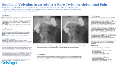
The patient had a history of type 2 diabetes complicated by foot osteomyelitis, atrial fibrillation, gastroesophageal reflux disease, and obstructive sleep apnea. Physical examination was notable for a soft abdomen with left upper quadrant tenerness tenderness. Initial serology showed an elevated white blood cell count, mild anemia, and elevated blood glucose levels. CT imaging with oral contrast demonstrated duodenal volvulus, characterized by obstruction and marked upstream dilation. The duodenum did not cross the midline, consistent with chronic malrotation. Additional findings included thickening of the esophageal wall, fatty replacement of the pancreas with calcification, and increased colonic stool burden.
The patient had a recent admission for right toe osteomyelitis, during which abdominal pain related to chronic malrotation had been observed. The patient had responded well to nasogastric tube placement and resumed oral intake prior to discharge.
Gastroenterology was consulted in which Esophagogastroduodenoscopy (EGD) revealed a dilated first portion of the duodenum and a volvulus in the third portion. Attempts to reduce the volvulus using endoscopy were unsuccessful, and no ischemia were observed. The patient subsequently underwent laparoscopy, during which internal hernia reduction, malrotation correction, and appendectomy were performed. The surgical intervention aimed to alleviate the duodenal obstruction and prevent future episodes of volvulus which was successful.
Discussion: This rare presentation of a duodenal volvulus secondary to intestinal malrotation in an elderly patient highlights the importance of considering undetected intestinal malrotation as a cause of small bowel volvulus. This case presentation has unique features including the age at presentation, as well as endoscopic evaluation for malignancy surveillance and possible therapeutic effect.

Disclosures:
Shiva F. Naidoo, MD1, Mahdi Taye, BA2, Hamzah Shariff, BA2, Lynzi Smith, BSc2, Sriram F. Chowdary, MD3, Divey Manocha, MD3. P1279 - Duodenal Volvulus in an Adult: A Rare Twist on Abdominal Pain, ACG 2023 Annual Scientific Meeting Abstracts. Vancouver, BC, Canada: American College of Gastroenterology.
1Geisinger Health System, Wilkes-Barre, PA; 2Geisinger Commonwealth School of Medicine, Scranton, PA; 3Geisinger Health System, Scranton, PA
Introduction: Midgut volvulus in adults is a rare encounter, due to the infrequency of this congenital anomaly and its early presentation. The reported incidence in this population range from 0.00001% to 0.19%. Majority (90%) of cases are diagnosed in infancy, with adult diagnoses comprising only 0.2-0.5%, and only a fraction of these presenting with midgut volvulus. As of 2018, there are only 92 reported cases of adults presenting with midgut volvulus with an average age of 40 years old. We present a case of a 79-year-old patient who was diagnosed with duodenal volvulus secondary to intestinal malrotation.
Case Description/Methods:
The patient had a history of type 2 diabetes complicated by foot osteomyelitis, atrial fibrillation, gastroesophageal reflux disease, and obstructive sleep apnea. Physical examination was notable for a soft abdomen with left upper quadrant tenerness tenderness. Initial serology showed an elevated white blood cell count, mild anemia, and elevated blood glucose levels. CT imaging with oral contrast demonstrated duodenal volvulus, characterized by obstruction and marked upstream dilation. The duodenum did not cross the midline, consistent with chronic malrotation. Additional findings included thickening of the esophageal wall, fatty replacement of the pancreas with calcification, and increased colonic stool burden.
The patient had a recent admission for right toe osteomyelitis, during which abdominal pain related to chronic malrotation had been observed. The patient had responded well to nasogastric tube placement and resumed oral intake prior to discharge.
Gastroenterology was consulted in which Esophagogastroduodenoscopy (EGD) revealed a dilated first portion of the duodenum and a volvulus in the third portion. Attempts to reduce the volvulus using endoscopy were unsuccessful, and no ischemia were observed. The patient subsequently underwent laparoscopy, during which internal hernia reduction, malrotation correction, and appendectomy were performed. The surgical intervention aimed to alleviate the duodenal obstruction and prevent future episodes of volvulus which was successful.
Discussion: This rare presentation of a duodenal volvulus secondary to intestinal malrotation in an elderly patient highlights the importance of considering undetected intestinal malrotation as a cause of small bowel volvulus. This case presentation has unique features including the age at presentation, as well as endoscopic evaluation for malignancy surveillance and possible therapeutic effect.

Figure: Figure 1. Small bowel follow through with oral contrast demonstrating volvulus at D2/D3 portion of duodenum
Disclosures:
Shiva Naidoo indicated no relevant financial relationships.
Mahdi Taye indicated no relevant financial relationships.
Hamzah Shariff indicated no relevant financial relationships.
Lynzi Smith indicated no relevant financial relationships.
Sriram Chowdary indicated no relevant financial relationships.
Divey Manocha indicated no relevant financial relationships.
Shiva F. Naidoo, MD1, Mahdi Taye, BA2, Hamzah Shariff, BA2, Lynzi Smith, BSc2, Sriram F. Chowdary, MD3, Divey Manocha, MD3. P1279 - Duodenal Volvulus in an Adult: A Rare Twist on Abdominal Pain, ACG 2023 Annual Scientific Meeting Abstracts. Vancouver, BC, Canada: American College of Gastroenterology.
