Sunday Poster Session
Category: Small Intestine
P1291 - Adenomyoma of the Ileum: A Rare Etiology of Abdominopelvic Pain
Sunday, October 22, 2023
3:30 PM - 7:00 PM PT
Location: Exhibit Hall

Has Audio
- LB
Lara Boudreaux, MD
LSU Health Sciences Center
New Orleans, LA
Presenting Author(s)
Jason Stibbe, MD, MS1, Stephen Wayne. Landreneau, MD2, Lara Boudreaux, MD3
1LSU Health, New Orleans, LA; 2Louisiana State University, New Orleans, LA; 3LSU Health Sciences Center, New Orleans, LA
Introduction: Adenomyoma is infrequently included in the typical differential of subepithelial small bowel lesions, owing to its rare occurrence and relative paucity of literature. Involvement of the ileum is an even rarer subset of these patients for which only case reports and series exist.
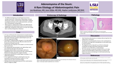
Case Description/Methods: We present a case of a 47 year-old woman with remote histories of endometriosis, hysterectomy, and ovarian cysts following with Gynecology for intermittent left lower quadrant abdominopelvic pain. She underwent contrasted CT Abdomen/Pelvis and was referred to our GI service after a 1.4cm left anterior pelvic soft tissue lesion was found with radiologic difficulty distinguishing bowel involvement (A). Colonoscopy including inspection of the distal 5 cm of terminal ileum was normal. Video capsule endoscopy (VCE) subsequently demonstrated a non-obstructing, round mass with a focal red spot (B) in the ileum. Retrograde double-balloon enteroscopy (DBE) confirmed a subepithelial mass in the mid ileum (C), whereby surface biopsies were obtained and a tattoo was placed distally using India Ink. The patient was referred for surgical resection and the mucosal biopsies demonstrated unremarkable overlying mucosa. Laparoscopy identified the marked region including a 1.4 cm subserosal projection. A 3-cm segment of mid ileum containing the mass was resected successfully and opened to reveal a 1.1cm submucosal mass with uninvolved margins. Histology demonstrated adenomyoma of the small intestine without malignancy.
Discussion: Fortunately, small intestinal adenomyoma is considered a benign entity arising from the submucosa or muscularis. However, diagnosis of this rare lesion can be challenging due to lack of characteristic radiographic imaging. Symptoms of non-periampullary lesions are also non-specific and can range from asymptomatic to vague intermittent pain to intussusception. Multi-modal small bowel endoscopy is therefore indispensable in the workup and treatment of these masses. As is often the case with subepithelial lesions, mucosal surface biopsies are unrevealing. Direct endoscopic visualization can narrow the differential but not always exclude more common mimickers such as GIST, NET, or endometrioma (in compatible history setting). VCE and DBE were able to confirm and pinpoint location of a mass in this case for a minimal, small bowel-preserving surgical resection. This case bolsters the literature including rare deep enteroscopic images to aid gastroenterologists in diagnostic awareness.

Disclosures:
Jason Stibbe, MD, MS1, Stephen Wayne. Landreneau, MD2, Lara Boudreaux, MD3. P1291 - Adenomyoma of the Ileum: A Rare Etiology of Abdominopelvic Pain, ACG 2023 Annual Scientific Meeting Abstracts. Vancouver, BC, Canada: American College of Gastroenterology.
1LSU Health, New Orleans, LA; 2Louisiana State University, New Orleans, LA; 3LSU Health Sciences Center, New Orleans, LA
Introduction: Adenomyoma is infrequently included in the typical differential of subepithelial small bowel lesions, owing to its rare occurrence and relative paucity of literature. Involvement of the ileum is an even rarer subset of these patients for which only case reports and series exist.
Case Description/Methods: We present a case of a 47 year-old woman with remote histories of endometriosis, hysterectomy, and ovarian cysts following with Gynecology for intermittent left lower quadrant abdominopelvic pain. She underwent contrasted CT Abdomen/Pelvis and was referred to our GI service after a 1.4cm left anterior pelvic soft tissue lesion was found with radiologic difficulty distinguishing bowel involvement (A). Colonoscopy including inspection of the distal 5 cm of terminal ileum was normal. Video capsule endoscopy (VCE) subsequently demonstrated a non-obstructing, round mass with a focal red spot (B) in the ileum. Retrograde double-balloon enteroscopy (DBE) confirmed a subepithelial mass in the mid ileum (C), whereby surface biopsies were obtained and a tattoo was placed distally using India Ink. The patient was referred for surgical resection and the mucosal biopsies demonstrated unremarkable overlying mucosa. Laparoscopy identified the marked region including a 1.4 cm subserosal projection. A 3-cm segment of mid ileum containing the mass was resected successfully and opened to reveal a 1.1cm submucosal mass with uninvolved margins. Histology demonstrated adenomyoma of the small intestine without malignancy.
Discussion: Fortunately, small intestinal adenomyoma is considered a benign entity arising from the submucosa or muscularis. However, diagnosis of this rare lesion can be challenging due to lack of characteristic radiographic imaging. Symptoms of non-periampullary lesions are also non-specific and can range from asymptomatic to vague intermittent pain to intussusception. Multi-modal small bowel endoscopy is therefore indispensable in the workup and treatment of these masses. As is often the case with subepithelial lesions, mucosal surface biopsies are unrevealing. Direct endoscopic visualization can narrow the differential but not always exclude more common mimickers such as GIST, NET, or endometrioma (in compatible history setting). VCE and DBE were able to confirm and pinpoint location of a mass in this case for a minimal, small bowel-preserving surgical resection. This case bolsters the literature including rare deep enteroscopic images to aid gastroenterologists in diagnostic awareness.

Figure: (A) Left anterior pelvic soft tissue density on contrasted CT Abdomen/Pelvis. (B) Small bowel mass with focal red spot on video capsule endoscopy. (C) Subepithelial mass in the mid ileum on retrograde double-balloon enteroscopy.
Disclosures:
Jason Stibbe indicated no relevant financial relationships.
Stephen Landreneau indicated no relevant financial relationships.
Lara Boudreaux indicated no relevant financial relationships.
Jason Stibbe, MD, MS1, Stephen Wayne. Landreneau, MD2, Lara Boudreaux, MD3. P1291 - Adenomyoma of the Ileum: A Rare Etiology of Abdominopelvic Pain, ACG 2023 Annual Scientific Meeting Abstracts. Vancouver, BC, Canada: American College of Gastroenterology.
