Sunday Poster Session
Category: Stomach
P1348 - Beyond the "Pale": Invasive Gastric Adenocarcinoma Hiding in Plain Sight
Sunday, October 22, 2023
3:30 PM - 7:00 PM PT
Location: Exhibit Hall

Has Audio

Aidan J. David
Case Western Reserve University
Cleveland, OH
Presenting Author(s)
Aidan J. David, 1, Brian M.. Fung, MD2, Joseph David, MD2
1Case Western Reserve University, Cleveland, OH; 2University of Arizona College of Medicine, Phoenix, AZ
Introduction: Mutations in the CDH1 gene, a tumor suppressor gene, are known to be responsible for the development of hereditary diffuse gastric cancer (HDGC). This form of gastric cancer often displays an infiltrative growth pattern, and is thus associated with a poor prognosis. Mucosal pale areas are the endoscopic abnormality most strongly associated with signet ring cell carcinoma (SRCC) foci. We describe a case of SRCC presenting as a diminutive area of slightly pale gastric mucosa in a patient with a CDH1 gene mutation who declined prophylactic gastrectomy.
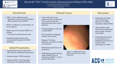
Case Description/Methods: A 49-year-old asymptomatic Caucasian female with a history of bilateral breast cancer underwent genetic testing which revealed a mutation of the CDH1 gene. She denied any family history of gastrointestinal malignancy. Her surgeon recommended prophylactic total gastrectomy given her elevated risk of gastric cancer. As she was reluctant to undergo prophylactic surgery, she presented to the gastroenterology clinic to discuss screening upper endoscopy. An esophagogastroduodenoscopy with mapping biopsies of the gastric mucosa was scheduled. On exam, her entire gastric mucosa was normal except for an inconspicuous, slightly pale area approximately 7mm in diameter on the mucosa of the stomach body (Figure 1). Two targeted biopsies of this area revealed SRCC foci, while mapping biopsies of the remainder of the stomach were normal. A staging CT of the chest, abdomen, and pelvis did not reveal any tumor spread, and a total gastrectomy was subsequently performed. The gastrectomy specimen was remarkable for a single residual focus of intramucosal SRCC 0.1 cm in greatest dimension.
Discussion: The goal of endoscopic screening or surveillance in patients with CDH1 gene mutations is to detect early SRCC. Such lesions can present with pale areas of the gastric mucosa endoscopically, but also with erosions, nodules, or polyps. If patients choose endoscopic surveillance over prophylactic gastrectomy, it is incumbent on the endoscopist to be prepared to take both standardized mapping biopsies, but also targeted biopsies of potentially suspicious lesions.

Disclosures:
Aidan J. David, 1, Brian M.. Fung, MD2, Joseph David, MD2. P1348 - Beyond the "Pale": Invasive Gastric Adenocarcinoma Hiding in Plain Sight, ACG 2023 Annual Scientific Meeting Abstracts. Vancouver, BC, Canada: American College of Gastroenterology.
1Case Western Reserve University, Cleveland, OH; 2University of Arizona College of Medicine, Phoenix, AZ
Introduction: Mutations in the CDH1 gene, a tumor suppressor gene, are known to be responsible for the development of hereditary diffuse gastric cancer (HDGC). This form of gastric cancer often displays an infiltrative growth pattern, and is thus associated with a poor prognosis. Mucosal pale areas are the endoscopic abnormality most strongly associated with signet ring cell carcinoma (SRCC) foci. We describe a case of SRCC presenting as a diminutive area of slightly pale gastric mucosa in a patient with a CDH1 gene mutation who declined prophylactic gastrectomy.
Case Description/Methods: A 49-year-old asymptomatic Caucasian female with a history of bilateral breast cancer underwent genetic testing which revealed a mutation of the CDH1 gene. She denied any family history of gastrointestinal malignancy. Her surgeon recommended prophylactic total gastrectomy given her elevated risk of gastric cancer. As she was reluctant to undergo prophylactic surgery, she presented to the gastroenterology clinic to discuss screening upper endoscopy. An esophagogastroduodenoscopy with mapping biopsies of the gastric mucosa was scheduled. On exam, her entire gastric mucosa was normal except for an inconspicuous, slightly pale area approximately 7mm in diameter on the mucosa of the stomach body (Figure 1). Two targeted biopsies of this area revealed SRCC foci, while mapping biopsies of the remainder of the stomach were normal. A staging CT of the chest, abdomen, and pelvis did not reveal any tumor spread, and a total gastrectomy was subsequently performed. The gastrectomy specimen was remarkable for a single residual focus of intramucosal SRCC 0.1 cm in greatest dimension.
Discussion: The goal of endoscopic screening or surveillance in patients with CDH1 gene mutations is to detect early SRCC. Such lesions can present with pale areas of the gastric mucosa endoscopically, but also with erosions, nodules, or polyps. If patients choose endoscopic surveillance over prophylactic gastrectomy, it is incumbent on the endoscopist to be prepared to take both standardized mapping biopsies, but also targeted biopsies of potentially suspicious lesions.

Figure: Figure 1. A slightly pale area of gastric mucosa found on upper endoscopy.
Disclosures:
Aidan David indicated no relevant financial relationships.
Brian Fung indicated no relevant financial relationships.
Joseph David indicated no relevant financial relationships.
Aidan J. David, 1, Brian M.. Fung, MD2, Joseph David, MD2. P1348 - Beyond the "Pale": Invasive Gastric Adenocarcinoma Hiding in Plain Sight, ACG 2023 Annual Scientific Meeting Abstracts. Vancouver, BC, Canada: American College of Gastroenterology.

