Sunday Poster Session
Category: Stomach
P1364 - Oxyntic Gland Adenoma: A Rare Finding During Evaluation of Iron Deficiency Anemia
Sunday, October 22, 2023
3:30 PM - 7:00 PM PT
Location: Exhibit Hall

Has Audio
- TG
Tyler Grantham, MD
Staten Island University Hospital
Staten Island, NY
Presenting Author(s)
Rajarajeshwari Ramachandran, MD1, Tyler Grantham, MD2, Vikash Kumar, MD1, Heidi Budke, MD3, Madhavi Reddy, MD1, Vinaya Gaduputi, MD3
1Brooklyn Hospital Center, Brooklyn, NY; 2Staten Island University Hospital, Staten Island, NY; 3Blanchard Valley Health System, Findlay, OH
Introduction: Oxyntic gland adenomas comprise less than 0.5% of all gastric tumors. They are composed of gland forming epithelial cells with predominantly chief cell differentiation resembling oxyntic glands. We are reporting a 22-year-old woman who was incidentally diagnosed with oxyntic gland adenoma during evaluation of iron deficiency anemia and successfully underwent endoscopic resection.
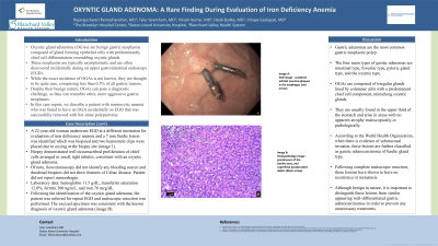
Case Description/Methods: A 22-year-old woman underwent esophagogastroduodenoscopy (EGD) at a different institution for evaluation of iron deficiency anemia and a 7 mm fundic lesion was identified which was biopsied and two hemostatic clips were placed due to oozing at the biopsy site. Biopsy demonstrated well circumscribed proliferation of chief cells arranged in small, tight tubules, consistent with an oxyntic gland adenoma. Of note, ileocolonoscopy did not identify any bleeding source and duodenal biopsies did not show features of Celiac disease. Patient did not report menorrhagia. Laboratory data: hemoglobin 11.5 g/dL, transferrin saturation 12.8%, ferritin 200 ng/mL, and iron 76 mcg/dL. Following the identification of the oxyntic gland adenoma, the patient was referred for repeat EGD. During the procedure, the oxyntic gland adenoma (image A) was identified endoscopic resection was performed. The excised specimen was consistent with the known diagnosis of oxyntic gland adenoma (image B).
Discussion: The four main types of gastric adenomas are intestinal type, foveolar type, pyloric gland type, and oxyntic type. Oxyntic gland adenomas are usually asymptomatic and found in the upper third of the stomach. Oxyntic gland adenomas without submucosal invasion have a benign behavior.

Disclosures:
Rajarajeshwari Ramachandran, MD1, Tyler Grantham, MD2, Vikash Kumar, MD1, Heidi Budke, MD3, Madhavi Reddy, MD1, Vinaya Gaduputi, MD3. P1364 - Oxyntic Gland Adenoma: A Rare Finding During Evaluation of Iron Deficiency Anemia, ACG 2023 Annual Scientific Meeting Abstracts. Vancouver, BC, Canada: American College of Gastroenterology.
1Brooklyn Hospital Center, Brooklyn, NY; 2Staten Island University Hospital, Staten Island, NY; 3Blanchard Valley Health System, Findlay, OH
Introduction: Oxyntic gland adenomas comprise less than 0.5% of all gastric tumors. They are composed of gland forming epithelial cells with predominantly chief cell differentiation resembling oxyntic glands. We are reporting a 22-year-old woman who was incidentally diagnosed with oxyntic gland adenoma during evaluation of iron deficiency anemia and successfully underwent endoscopic resection.
Case Description/Methods: A 22-year-old woman underwent esophagogastroduodenoscopy (EGD) at a different institution for evaluation of iron deficiency anemia and a 7 mm fundic lesion was identified which was biopsied and two hemostatic clips were placed due to oozing at the biopsy site. Biopsy demonstrated well circumscribed proliferation of chief cells arranged in small, tight tubules, consistent with an oxyntic gland adenoma. Of note, ileocolonoscopy did not identify any bleeding source and duodenal biopsies did not show features of Celiac disease. Patient did not report menorrhagia. Laboratory data: hemoglobin 11.5 g/dL, transferrin saturation 12.8%, ferritin 200 ng/mL, and iron 76 mcg/dL. Following the identification of the oxyntic gland adenoma, the patient was referred for repeat EGD. During the procedure, the oxyntic gland adenoma (image A) was identified endoscopic resection was performed. The excised specimen was consistent with the known diagnosis of oxyntic gland adenoma (image B).
Discussion: The four main types of gastric adenomas are intestinal type, foveolar type, pyloric gland type, and oxyntic type. Oxyntic gland adenomas are usually asymptomatic and found in the upper third of the stomach. Oxyntic gland adenomas without submucosal invasion have a benign behavior.

Figure: Image A: EGD image - oxyntic gland adenoma with hemostatic clips placed during previous EGD (red arrow)
Image B: Histopathology image - well circumscribed proliferation of chief cells arranged in small, tight tubules
Image B: Histopathology image - well circumscribed proliferation of chief cells arranged in small, tight tubules
Disclosures:
Rajarajeshwari Ramachandran indicated no relevant financial relationships.
Tyler Grantham indicated no relevant financial relationships.
Vikash Kumar indicated no relevant financial relationships.
Heidi Budke indicated no relevant financial relationships.
Madhavi Reddy indicated no relevant financial relationships.
Vinaya Gaduputi indicated no relevant financial relationships.
Rajarajeshwari Ramachandran, MD1, Tyler Grantham, MD2, Vikash Kumar, MD1, Heidi Budke, MD3, Madhavi Reddy, MD1, Vinaya Gaduputi, MD3. P1364 - Oxyntic Gland Adenoma: A Rare Finding During Evaluation of Iron Deficiency Anemia, ACG 2023 Annual Scientific Meeting Abstracts. Vancouver, BC, Canada: American College of Gastroenterology.
