Sunday Poster Session
Category: Stomach
P1394 - Gastrointestinal Spindle Cell Neoplasms: A 2-Case Study of Resected Low-Grade GIST and an Ileal Leiomyoma With Clinico-Pathological Correlation
Sunday, October 22, 2023
3:30 PM - 7:00 PM PT
Location: Exhibit Hall

Has Audio

Lisa Centeno, MD
University of Santo Tomas Faculty of Medicine and Surgery
FRESNO, CA
Presenting Author(s)
Lisa Centeno, MD1, Amirah Salem, BS2, Yasser H. Salem, MD3
1University of Santo Tomas Faculty of Medicine and Surgery, Manila, National Capital Region, Philippines; 2University of California, Irvine, Irvine, CA; 3South Coast Global Medical Center, Santa Ana, CA
Introduction: Gastrointestinal stromal tumors (GISTs) arise from the interstitial cells of Cajal, while leiomyomas are muscularis propria or muscularis mucosa derivatives. With differing malignant potentials and immunohistochemical profiles, both can be assessed as spindle cell neoplasms on frozen section. Resection of both tumors depends on tumor location and size, extent of spread, as well as clinical presentation and confidence in preoperative diagnosis. We present cases of a low-grade GIST resected by partial gastrectomy, and of an ileal leiomyoma resected by open laparotomy.
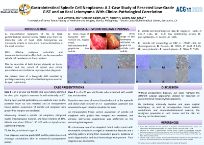
Case Description/Methods: Case 1 is of a 68 year old female who presented with abdominal pain and gastric mass on CT. Partial gastrectomy was done, and an intraoperative frozen section assessment of spindle cell neoplasm with grossly free margins was rendered. The singular 4x3.5x3 cm, roughly ovoid, well-circumscribed grayish-tan firm nodule. Microscopy showed spindle cell neoplasm, elongated nuclei, inconspicuous nucleoli, and focal necrosis of 10%. Mitosis was about 3/5mm2, 50 HPF, and DOG1 and CD117 C-kit were positive, and S100, pancytokeratin, desmin and SMA were negative. Ki-67 showed low-proliferating activity, Final diagnosis was low-grade GIST, and the patient received oncology consultations after an uneventful postoperative period.
Case 2 is of a 25 year old female who presented with weight loss and diarrhea. Resection of a mass found adjacent to the appendix and distal small on CT was done. An intraoperative frozen section assessment of spindle cell neoplasm with grossly free margins was rendered, and primary, side-to-side anastomosis was performed on the remaining ileal segment. The 4x0.5x4x3 cm, grayish-tan partially cystic nodule. On microscopy, round to elongated, blunt-ended nuclei with eosinophilic cytoplasm arranged as intersection fascicles and a whorling pattern arising from muscularis propria. Evidence of cystic degeneration and focal hemorrhage were present. Desmin and SMA, and CD117, DOG1, S100, chromogranin A and synaptophysin were negative. Ki-67 proliferation index was 3-5%. Final diagnosis was leiomyoma.
Discussion: Without preoperative biopsies, our cases highlight the different surgical approaches utilized for resection of gastrointestinal mesenchymal neoplasms. By combining minimally invasive, open surgical techniques, as well as intraoperative frozen section consultation and immunohistochemical analysis, the malignant properties of such tumors and plan for therapy can be determined.

Disclosures:
Lisa Centeno, MD1, Amirah Salem, BS2, Yasser H. Salem, MD3. P1394 - Gastrointestinal Spindle Cell Neoplasms: A 2-Case Study of Resected Low-Grade GIST and an Ileal Leiomyoma With Clinico-Pathological Correlation, ACG 2023 Annual Scientific Meeting Abstracts. Vancouver, BC, Canada: American College of Gastroenterology.
1University of Santo Tomas Faculty of Medicine and Surgery, Manila, National Capital Region, Philippines; 2University of California, Irvine, Irvine, CA; 3South Coast Global Medical Center, Santa Ana, CA
Introduction: Gastrointestinal stromal tumors (GISTs) arise from the interstitial cells of Cajal, while leiomyomas are muscularis propria or muscularis mucosa derivatives. With differing malignant potentials and immunohistochemical profiles, both can be assessed as spindle cell neoplasms on frozen section. Resection of both tumors depends on tumor location and size, extent of spread, as well as clinical presentation and confidence in preoperative diagnosis. We present cases of a low-grade GIST resected by partial gastrectomy, and of an ileal leiomyoma resected by open laparotomy.
Case Description/Methods: Case 1 is of a 68 year old female who presented with abdominal pain and gastric mass on CT. Partial gastrectomy was done, and an intraoperative frozen section assessment of spindle cell neoplasm with grossly free margins was rendered. The singular 4x3.5x3 cm, roughly ovoid, well-circumscribed grayish-tan firm nodule. Microscopy showed spindle cell neoplasm, elongated nuclei, inconspicuous nucleoli, and focal necrosis of 10%. Mitosis was about 3/5mm2, 50 HPF, and DOG1 and CD117 C-kit were positive, and S100, pancytokeratin, desmin and SMA were negative. Ki-67 showed low-proliferating activity, Final diagnosis was low-grade GIST, and the patient received oncology consultations after an uneventful postoperative period.
Case 2 is of a 25 year old female who presented with weight loss and diarrhea. Resection of a mass found adjacent to the appendix and distal small on CT was done. An intraoperative frozen section assessment of spindle cell neoplasm with grossly free margins was rendered, and primary, side-to-side anastomosis was performed on the remaining ileal segment. The 4x0.5x4x3 cm, grayish-tan partially cystic nodule. On microscopy, round to elongated, blunt-ended nuclei with eosinophilic cytoplasm arranged as intersection fascicles and a whorling pattern arising from muscularis propria. Evidence of cystic degeneration and focal hemorrhage were present. Desmin and SMA, and CD117, DOG1, S100, chromogranin A and synaptophysin were negative. Ki-67 proliferation index was 3-5%. Final diagnosis was leiomyoma.
Discussion: Without preoperative biopsies, our cases highlight the different surgical approaches utilized for resection of gastrointestinal mesenchymal neoplasms. By combining minimally invasive, open surgical techniques, as well as intraoperative frozen section consultation and immunohistochemical analysis, the malignant properties of such tumors and plan for therapy can be determined.

Figure: Top row (left to right): gross image of low-grade GIST, spindle cell morphology on H and E, Dog1+, CD117 c-kit+, Ki-67 low proliferating activity.
Bottom row (left to right): gross image of leiomyoma, spindle cell morphology on H and E, Desmin+, SMA+, Ki-67 of 3-5%
Bottom row (left to right): gross image of leiomyoma, spindle cell morphology on H and E, Desmin+, SMA+, Ki-67 of 3-5%
Disclosures:
Lisa Centeno indicated no relevant financial relationships.
Amirah Salem indicated no relevant financial relationships.
Yasser Salem indicated no relevant financial relationships.
Lisa Centeno, MD1, Amirah Salem, BS2, Yasser H. Salem, MD3. P1394 - Gastrointestinal Spindle Cell Neoplasms: A 2-Case Study of Resected Low-Grade GIST and an Ileal Leiomyoma With Clinico-Pathological Correlation, ACG 2023 Annual Scientific Meeting Abstracts. Vancouver, BC, Canada: American College of Gastroenterology.
