Sunday Poster Session
Category: Stomach
P1397 - An Uncommon Case of Pancreatic Cancer Metastasizing to the Stomach
Sunday, October 22, 2023
3:30 PM - 7:00 PM PT
Location: Exhibit Hall

Has Audio
- HE
Henry Egbuchiem, Bsc, MD
University Hospitals Geauga Medical Center
Chardon, OH
Presenting Author(s)
Henry Egbuchiem, Bsc, MD1, Alwatheq Alitelat, 1, Joseph Amoah, MD1, Nkemputaife Onyechi, MD2, Suhani Dalal, MD1, Mohammad Mazumder, MD, FACG2, Parvez Khambatta, MD, FACG1
1University Hospitals Geauga Medical Center, Chardon, OH; 2University Hospitals, Chardon, OH
Introduction: The incidence of gastric metastases from pancreatic cancer is unknown due to the low sensitivity of imaging studies, which makes clinical detection difficult. However, gastric malignancy is the fourth most common cancer worldwide, with an estimated 1.1 million new cases and 800000 deaths in 2020. Gastric cancer secondary to Pancreatic metastasis is a rare condition. Current literature stipulates that pancreatic cancer metastasizes mostly through lymphatics to surrounding structures. While it is common for gastric cancer to metastasize to the pancreas, it is uncommon for pancreatic metastasis to go from the pancreas to the stomach. In this case, we described a case of an 89-year-old female with Stage IV pancreatic cancer that presented with a Malignant gastric ulcer found on endoscopy and after doing a biopsy.
Case Description/Methods: An 89-year-old female with a history of unprovoked PE (on Eliquis) hypothyroidism, HTN, essential tremors, Stage IV pancreatic cancer pancreatic body/tail, with an uncharacterized liver lesion, Ca 19-9 positive >4000-not currently on chemotherapy.
She presented to the hospital due to a 2weeks history of weakness, non-bloody diarrhea, and fever. Vital signals are unremarkable. On general examination, she looked frail & had conjunctival pallor. CVS is significant for ejection systolic murmur. Labs showed hemoglobin of 4.5, INR of 3.1, Albumin 2.9, Total protein was 5.8, Ca 7.8, Cl of 111, bicarb of 19, BUN 35, Cr 1.16 & a positive FOBT.
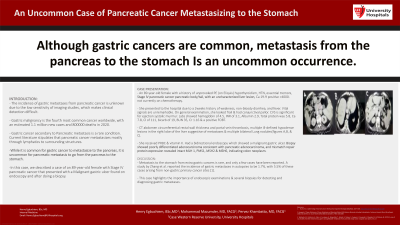
CT abdomen circumferential rectal wall thickness and portal vein thrombosis, multiple ill-defined hypodense lesions in the right lobe of the liver suggestive of metastases & multiple bilateral Lung nodules(figures A,B, & C). She received PRBC & vitamin K. Had a bidirectional endoscopy which showed a malignant gastric ulcer. Biopsy showed poorly differentiated adenocarcinoma consistent with pancreatic adenocarcinoma, and mismatch repair protein expression revealed intact MLH 1, PMS1, MSH2 & MSH6, indicating colon neoplasm.
Discussion: Metastasis to the stomach from extra gastric cancers is rare, and only a few cases have been reported. A study by Zhang et al. reported the incidence of gastric metastases in autopsies to be 1.7%, with 5.5% of these cases arising from non-gastric primary cancer sites [7]. This case highlights the importance of endoscopic examinations & several biopsies for detecting and diagnosing gastric metastases.

Disclosures:
Henry Egbuchiem, Bsc, MD1, Alwatheq Alitelat, 1, Joseph Amoah, MD1, Nkemputaife Onyechi, MD2, Suhani Dalal, MD1, Mohammad Mazumder, MD, FACG2, Parvez Khambatta, MD, FACG1. P1397 - An Uncommon Case of Pancreatic Cancer Metastasizing to the Stomach, ACG 2023 Annual Scientific Meeting Abstracts. Vancouver, BC, Canada: American College of Gastroenterology.
1University Hospitals Geauga Medical Center, Chardon, OH; 2University Hospitals, Chardon, OH
Introduction: The incidence of gastric metastases from pancreatic cancer is unknown due to the low sensitivity of imaging studies, which makes clinical detection difficult. However, gastric malignancy is the fourth most common cancer worldwide, with an estimated 1.1 million new cases and 800000 deaths in 2020. Gastric cancer secondary to Pancreatic metastasis is a rare condition. Current literature stipulates that pancreatic cancer metastasizes mostly through lymphatics to surrounding structures. While it is common for gastric cancer to metastasize to the pancreas, it is uncommon for pancreatic metastasis to go from the pancreas to the stomach. In this case, we described a case of an 89-year-old female with Stage IV pancreatic cancer that presented with a Malignant gastric ulcer found on endoscopy and after doing a biopsy.
Case Description/Methods: An 89-year-old female with a history of unprovoked PE (on Eliquis) hypothyroidism, HTN, essential tremors, Stage IV pancreatic cancer pancreatic body/tail, with an uncharacterized liver lesion, Ca 19-9 positive >4000-not currently on chemotherapy.
She presented to the hospital due to a 2weeks history of weakness, non-bloody diarrhea, and fever. Vital signals are unremarkable. On general examination, she looked frail & had conjunctival pallor. CVS is significant for ejection systolic murmur. Labs showed hemoglobin of 4.5, INR of 3.1, Albumin 2.9, Total protein was 5.8, Ca 7.8, Cl of 111, bicarb of 19, BUN 35, Cr 1.16 & a positive FOBT.
CT abdomen circumferential rectal wall thickness and portal vein thrombosis, multiple ill-defined hypodense lesions in the right lobe of the liver suggestive of metastases & multiple bilateral Lung nodules(figures A,B, & C). She received PRBC & vitamin K. Had a bidirectional endoscopy which showed a malignant gastric ulcer. Biopsy showed poorly differentiated adenocarcinoma consistent with pancreatic adenocarcinoma, and mismatch repair protein expression revealed intact MLH 1, PMS1, MSH2 & MSH6, indicating colon neoplasm.
Discussion: Metastasis to the stomach from extra gastric cancers is rare, and only a few cases have been reported. A study by Zhang et al. reported the incidence of gastric metastases in autopsies to be 1.7%, with 5.5% of these cases arising from non-gastric primary cancer sites [7]. This case highlights the importance of endoscopic examinations & several biopsies for detecting and diagnosing gastric metastases.

Figure: figure A, B and C shows numerous hepatic lesions, Also pancreatic mass is evident in figure A
Disclosures:
Henry Egbuchiem indicated no relevant financial relationships.
Alwatheq Alitelat indicated no relevant financial relationships.
Joseph Amoah indicated no relevant financial relationships.
Nkemputaife Onyechi indicated no relevant financial relationships.
Suhani Dalal indicated no relevant financial relationships.
Mohammad Mazumder indicated no relevant financial relationships.
Parvez Khambatta indicated no relevant financial relationships.
Henry Egbuchiem, Bsc, MD1, Alwatheq Alitelat, 1, Joseph Amoah, MD1, Nkemputaife Onyechi, MD2, Suhani Dalal, MD1, Mohammad Mazumder, MD, FACG2, Parvez Khambatta, MD, FACG1. P1397 - An Uncommon Case of Pancreatic Cancer Metastasizing to the Stomach, ACG 2023 Annual Scientific Meeting Abstracts. Vancouver, BC, Canada: American College of Gastroenterology.
