Sunday Poster Session
Category: Stomach
P1404 - Intestinal Metaplasia Advancing to Gastric Adenocarcinoma Following Cardiac Transplantation
Sunday, October 22, 2023
3:30 PM - 7:00 PM PT
Location: Exhibit Hall

Has Audio
- CW
Ciara White
Henry Ford Heath System
Detroit, Michigan
Presenting Author(s)
Ciara White, 1, Adarsh Varma, MD2, Syed-Mohammed Jafri, MD3
1Henry Ford Heath System, Detroit, MI; 2Henry Ford Health, Detroit, MI; 3Henry Ford Health System, Detroit, MI
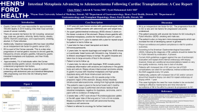
Introduction: Intestinal Metaplasia (IM) has been identified as a risk factor for gastric cancer (GC). We describe a case of a patient presenting with abdominal pain and Intestinal Metaplasia (IM) progressing over time into GC following heart transplant.
Case Description/Methods: A 57 year old male former smoker with gastroesophageal reflux disease (GERD) presents with abdominal pain and weight loss. An upper gastrointestinal endoscopy (EGD) shows 2 ulcers on the lesser curvature of the stomach. Biopsy shows gastritis with intestinal metaplasia, positive for helicobacter pylori (H. pylori). Triple therapy for H. pylori is started with a plan for repeat scope but is lost to follow up. 1 year later he has a heart transplant and starts immunosuppressants. 4 years later, he reports dysphagia and weight loss. EGD shows a 4 centimeter hiatal hernia with Schatzki’s ring. Schatzki’s ring is dilated; esophageal biopsy is benign. No gastric biopsies are taken. 1 year later he reports dysphagia and weight loss. EGD is initiated, but not completed due to food matter. 2 years later, he returns with dysphagia. EGD reveals patchy moderate inflammation with erosions and erythema in the gastric body and antrum. Biopsy shows intramucosal carcinoma arising in gastric mucosa with intestinal metaplasia and high-grade dysplasia along with focal intramucosal carcinoma. 1 month later, EGD shows a 20 mm sessile polyp in the prepyloric region of the stomach. Biopsy shows intestinal, intramucosal adenocarcinoma, arising from high-grade dysplasia. 3 months later, EGD shows residual focal intestinal metaplasia, negative for dysplasia, carcinoma, and H. pylori with no evidence of malignancy. 2 weeks later, a PET scan shows F-fluorodeoxyglucose uptake in the left upper lobe of the lung and a 7 mm x 9 mm nodule. Biopsy is positive for non-small cell carcinoma favoring squamous cell carcinoma. Patient is referred to oncology for further treatment.
Discussion: Intestinal Metaplasia (IM) is the transformation of stomach epithelium to intestinal epithelium. The progression of IM to GC is rare, occurring in 0.09% of cases. Risk factors include: H. pylori, family history of GC, GERD, Barett’s esophagus, acid suppressants, obesity and tobacco. Proper surveillance and patient compliance is vital. The American Gastroenterological Association Recommends repeat EGD with mucosal visualization and gastric biopsies of the antrum, body and any lesions every 3–5 years in patients with incidentally detected GIM.
Disclosures:
Ciara White, 1, Adarsh Varma, MD2, Syed-Mohammed Jafri, MD3. P1404 - Intestinal Metaplasia Advancing to Gastric Adenocarcinoma Following Cardiac Transplantation, ACG 2023 Annual Scientific Meeting Abstracts. Vancouver, BC, Canada: American College of Gastroenterology.
1Henry Ford Heath System, Detroit, MI; 2Henry Ford Health, Detroit, MI; 3Henry Ford Health System, Detroit, MI
Introduction: Intestinal Metaplasia (IM) has been identified as a risk factor for gastric cancer (GC). We describe a case of a patient presenting with abdominal pain and Intestinal Metaplasia (IM) progressing over time into GC following heart transplant.
Case Description/Methods: A 57 year old male former smoker with gastroesophageal reflux disease (GERD) presents with abdominal pain and weight loss. An upper gastrointestinal endoscopy (EGD) shows 2 ulcers on the lesser curvature of the stomach. Biopsy shows gastritis with intestinal metaplasia, positive for helicobacter pylori (H. pylori). Triple therapy for H. pylori is started with a plan for repeat scope but is lost to follow up. 1 year later he has a heart transplant and starts immunosuppressants. 4 years later, he reports dysphagia and weight loss. EGD shows a 4 centimeter hiatal hernia with Schatzki’s ring. Schatzki’s ring is dilated; esophageal biopsy is benign. No gastric biopsies are taken. 1 year later he reports dysphagia and weight loss. EGD is initiated, but not completed due to food matter. 2 years later, he returns with dysphagia. EGD reveals patchy moderate inflammation with erosions and erythema in the gastric body and antrum. Biopsy shows intramucosal carcinoma arising in gastric mucosa with intestinal metaplasia and high-grade dysplasia along with focal intramucosal carcinoma. 1 month later, EGD shows a 20 mm sessile polyp in the prepyloric region of the stomach. Biopsy shows intestinal, intramucosal adenocarcinoma, arising from high-grade dysplasia. 3 months later, EGD shows residual focal intestinal metaplasia, negative for dysplasia, carcinoma, and H. pylori with no evidence of malignancy. 2 weeks later, a PET scan shows F-fluorodeoxyglucose uptake in the left upper lobe of the lung and a 7 mm x 9 mm nodule. Biopsy is positive for non-small cell carcinoma favoring squamous cell carcinoma. Patient is referred to oncology for further treatment.
Discussion: Intestinal Metaplasia (IM) is the transformation of stomach epithelium to intestinal epithelium. The progression of IM to GC is rare, occurring in 0.09% of cases. Risk factors include: H. pylori, family history of GC, GERD, Barett’s esophagus, acid suppressants, obesity and tobacco. Proper surveillance and patient compliance is vital. The American Gastroenterological Association Recommends repeat EGD with mucosal visualization and gastric biopsies of the antrum, body and any lesions every 3–5 years in patients with incidentally detected GIM.
Disclosures:
Ciara White indicated no relevant financial relationships.
Adarsh Varma indicated no relevant financial relationships.
Syed-Mohammed Jafri: Gilead, Takeda, Abbvie – Advisor or Review Panel Member, Speakers Bureau.
Ciara White, 1, Adarsh Varma, MD2, Syed-Mohammed Jafri, MD3. P1404 - Intestinal Metaplasia Advancing to Gastric Adenocarcinoma Following Cardiac Transplantation, ACG 2023 Annual Scientific Meeting Abstracts. Vancouver, BC, Canada: American College of Gastroenterology.
