Sunday Poster Session
Category: Stomach
P1405 - An Unexpected Connection: A Rare Case of Gastrosplenic Fistula in Diffuse Large B-Cell Lymphoma
Sunday, October 22, 2023
3:30 PM - 7:00 PM PT
Location: Exhibit Hall

Has Audio

Muna Okafor, MD
University Hospitals Case Medical Center, Case Western Reserve University
Cleveland, OH
Presenting Author(s)
Award: Presidential Poster Award
Muna Okafor, MD1, Dayyan Adoor, MD1, Brooke Glessing, MD2, Gregory Cooper, MD3
1University Hospitals Case Medical Center, Case Western Reserve University, Cleveland, OH; 2Digestive Health Institute, University Hospitals Cleveland Medical Center, Cleveland, OH; 3Case Western Reserve/UHCMC, Cleveland, OH
Introduction: Gastrosplenic fistula (GSF) is a rare condition, often arising as a complication of gastric or splenic malignancies. Diagnosis and management can prove challenging due to nonspecific symptoms, and requires prompt identification to prevent serious complications. Here, we present a rare case of gastrosplenic fistula secondary to diffuse large B cell lymphoma in a patient who presented with chest and abdominal pain.
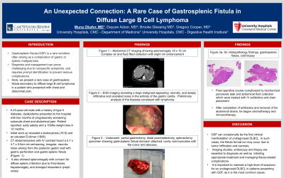
Case Description/Methods: A 43-year-old male with a history of type II diabetes, dyslipidemia presented to the hospital with subacute chest and abdominal pain. His chest pain started two months prior, described as progressively worsening sharp, stabbing and radiating to the back and left shoulder. He also reported epigastric and left upper quadrant pain that worsened after eating any solid foods. Patient endorsed early satiety and a 100lbs weight loss in 12 months. Initial work up revealed a leukocytosis (16.9) and an elevated D-dimer (1906). CT abdomen/pelvis with IV contrast found a 5.7 x 4.7 x 5.5cm rim-enhancing, irregular, necrotic mass arising from the posterior gastric wall with gastric perforation and gastro-splenic fistula (Fig. 1). It also showed splenomegaly with concern for diffuse splenic infarction due to thrombosis, hepatomegaly, and enlarged mesenteric lymph nodes. Upper endoscopy found a large, malignant-appearing, necrotic, and deeply infiltrative and ulcerated mass occupying the entirety of the gastric cardia (Fig. 2). Preliminary analysis of the biopsies was consistent with lymphoma.
He underwent partial gastrectomy, distal pancreatectomy, and splenectomy (Fig. 3). Final biopsies confirmed a new diagnosis of diffuse large B-cell lymphoma (Fig. 4a and 4b). His post-operative course was complicated by biochemical pancreatic leak and abdominal fluid collection which were treated with IV antibiotics and drain placement. After completion of antibiotics and removal of his abdominal drains, he began chemotherapy and immunotherapy.
Discussion: GSF can occasionally be the first clinical manifestation of undiagnosed DLBCL. In such cases, the fistula formation may occur due to tumor infiltration and necrosis. Imaging studies, endoscopy and biopsy are essential to diagnosis. This is crucial for initiating appropriate treatment and managing fistula-related complications. It is important to maintain a high level of suspicion for an undiagnosed DLBCL in patients presenting with GSF, as it is the most common cause.

Disclosures:
Muna Okafor, MD1, Dayyan Adoor, MD1, Brooke Glessing, MD2, Gregory Cooper, MD3. P1405 - An Unexpected Connection: A Rare Case of Gastrosplenic Fistula in Diffuse Large B-Cell Lymphoma, ACG 2023 Annual Scientific Meeting Abstracts. Vancouver, BC, Canada: American College of Gastroenterology.
Muna Okafor, MD1, Dayyan Adoor, MD1, Brooke Glessing, MD2, Gregory Cooper, MD3
1University Hospitals Case Medical Center, Case Western Reserve University, Cleveland, OH; 2Digestive Health Institute, University Hospitals Cleveland Medical Center, Cleveland, OH; 3Case Western Reserve/UHCMC, Cleveland, OH
Introduction: Gastrosplenic fistula (GSF) is a rare condition, often arising as a complication of gastric or splenic malignancies. Diagnosis and management can prove challenging due to nonspecific symptoms, and requires prompt identification to prevent serious complications. Here, we present a rare case of gastrosplenic fistula secondary to diffuse large B cell lymphoma in a patient who presented with chest and abdominal pain.
Case Description/Methods: A 43-year-old male with a history of type II diabetes, dyslipidemia presented to the hospital with subacute chest and abdominal pain. His chest pain started two months prior, described as progressively worsening sharp, stabbing and radiating to the back and left shoulder. He also reported epigastric and left upper quadrant pain that worsened after eating any solid foods. Patient endorsed early satiety and a 100lbs weight loss in 12 months. Initial work up revealed a leukocytosis (16.9) and an elevated D-dimer (1906). CT abdomen/pelvis with IV contrast found a 5.7 x 4.7 x 5.5cm rim-enhancing, irregular, necrotic mass arising from the posterior gastric wall with gastric perforation and gastro-splenic fistula (Fig. 1). It also showed splenomegaly with concern for diffuse splenic infarction due to thrombosis, hepatomegaly, and enlarged mesenteric lymph nodes. Upper endoscopy found a large, malignant-appearing, necrotic, and deeply infiltrative and ulcerated mass occupying the entirety of the gastric cardia (Fig. 2). Preliminary analysis of the biopsies was consistent with lymphoma.
He underwent partial gastrectomy, distal pancreatectomy, and splenectomy (Fig. 3). Final biopsies confirmed a new diagnosis of diffuse large B-cell lymphoma (Fig. 4a and 4b). His post-operative course was complicated by biochemical pancreatic leak and abdominal fluid collection which were treated with IV antibiotics and drain placement. After completion of antibiotics and removal of his abdominal drains, he began chemotherapy and immunotherapy.
Discussion: GSF can occasionally be the first clinical manifestation of undiagnosed DLBCL. In such cases, the fistula formation may occur due to tumor infiltration and necrosis. Imaging studies, endoscopy and biopsy are essential to diagnosis. This is crucial for initiating appropriate treatment and managing fistula-related complications. It is important to maintain a high level of suspicion for an undiagnosed DLBCL in patients presenting with GSF, as it is the most common cause.

Figure: Figure 1 – Abdominal CT imaging showing splenomegaly 18 x 15 cm
Complex air and fluid-filled collection with slight rim enhancement
Figure 2: EGD imaging revealing a large necrotic, and deeply infiltrative, and ulcerated mass in the entirety of the gastric cardia.
Figure 3 : specimen showing gastrosplenic fistula stomach attached; cavity communicates with the tumor and abscess.
Figure 4a, 4b: Histopathology findings, gastrosplenic fistula, cold biopsy
Disclosures:
Muna Okafor indicated no relevant financial relationships.
Dayyan Adoor indicated no relevant financial relationships.
Brooke Glessing indicated no relevant financial relationships.
Gregory Cooper indicated no relevant financial relationships.
Muna Okafor, MD1, Dayyan Adoor, MD1, Brooke Glessing, MD2, Gregory Cooper, MD3. P1405 - An Unexpected Connection: A Rare Case of Gastrosplenic Fistula in Diffuse Large B-Cell Lymphoma, ACG 2023 Annual Scientific Meeting Abstracts. Vancouver, BC, Canada: American College of Gastroenterology.

