Sunday Poster Session
Category: Stomach
P1406 - A Rare Case of Myeloid Sarcoma of the Stomach
Sunday, October 22, 2023
3:30 PM - 7:00 PM PT
Location: Exhibit Hall


Saraswathi Lakkasani, MD
Saint Michael's Medical Center, New York Medical College
Glen Allen, VA
Presenting Author(s)
Navjot Kaur Grewal, MD1, Saraswathi Lakkasani, MD2, Eliezer Seguerra, MD1, Yatinder Bains, MD3, Rewanth Katamreddy, MD4
1Saint Michael's Medical Center, Newark, NJ; 2Saint Michael's Medical Center, New York Medical College, Newark, NJ; 3Saint Michale's Medical Center, Newark, NJ; 4Saint Michael’s Medical Center, Newark, NJ
Introduction: Isolated myeloid sarcomas (MS) are proliferation of myeloblasts at extramedullary sites without any bone marrow involvement. These tumors are extremely rare and pose a diagnostic and therapeutic challenge.
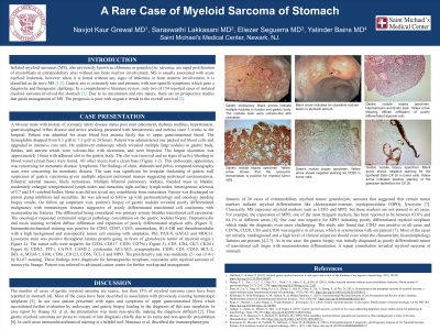
Case Description/Methods: A 68-year male with history of coronary artery disease and stent placement, diabetes mellitus, hypertension, gastroesophageal reflux disease and smoking presented with hematemesis and melena since 3 weeks. Patient was admitted for acute blood loss anemia due to upper gastrointestinal bleed. The hemoglobin dropped from 8.3 g/dl to 7.1 g/dl in 24 hours. Patient was transfused. He underwent endoscopy which revealed multiple large nodules in gastric body, fundus, and antrum with volcano-like with ulceration, which were biopsied (Figure1). This endoscopic appearance was concerning for metastatic disease/ lymphoma. Patient’s biopsy of gastric nodules revealed poorly differentiated malignancy with immunotypic features suggestive of transitional cell carcinoma with neuroendocrine features. The oncologist requested a second surgical pathology consultation. Haematoxylin and eosin staining revealed diffuse infiltration and expansion of poorly differentiated atypical cells. Immunohistochemical staining was positive for lysozyme stain throughout lamina propria going in favor of granulocyte tumor of myeloid origin. The tumor cells were negative for CD20, CD79a, CD3, CK20, CD30 and myeloperoxidase (MPO). These findings were diagnostic for hematopoietic neoplasm, consistent with myeloid sarcoma of monocytic lineage.
Discussion: Our patient presented with signs and symptoms of upper gastrointestinal bleed which necessitated further evaluation leading to the diagnosis of this rare neoplasm. In case report by Huang XL et al, the presentation was non-specific making the diagnosis difficult. In such cases immunohistochemical staining is helpful. Menasce et al, described the immunotypic features of 26 cases of extramedullary MS that suggested that tumor markers like MPO and lysozyme indicate myeloid differentiation. Classically, MS expresses myeloid markers but they are not present in all cases. Our case was negative for MPO indicating poorly differentiated myeloid neoplasm which made the diagnosis challenging. The study also found that CD79a, CD20, CD3 and CD30 were negative in all cases, which is synonymous with our case.

Disclosures:
Navjot Kaur Grewal, MD1, Saraswathi Lakkasani, MD2, Eliezer Seguerra, MD1, Yatinder Bains, MD3, Rewanth Katamreddy, MD4. P1406 - A Rare Case of Myeloid Sarcoma of the Stomach, ACG 2023 Annual Scientific Meeting Abstracts. Vancouver, BC, Canada: American College of Gastroenterology.
1Saint Michael's Medical Center, Newark, NJ; 2Saint Michael's Medical Center, New York Medical College, Newark, NJ; 3Saint Michale's Medical Center, Newark, NJ; 4Saint Michael’s Medical Center, Newark, NJ
Introduction: Isolated myeloid sarcomas (MS) are proliferation of myeloblasts at extramedullary sites without any bone marrow involvement. These tumors are extremely rare and pose a diagnostic and therapeutic challenge.
Case Description/Methods: A 68-year male with history of coronary artery disease and stent placement, diabetes mellitus, hypertension, gastroesophageal reflux disease and smoking presented with hematemesis and melena since 3 weeks. Patient was admitted for acute blood loss anemia due to upper gastrointestinal bleed. The hemoglobin dropped from 8.3 g/dl to 7.1 g/dl in 24 hours. Patient was transfused. He underwent endoscopy which revealed multiple large nodules in gastric body, fundus, and antrum with volcano-like with ulceration, which were biopsied (Figure1). This endoscopic appearance was concerning for metastatic disease/ lymphoma. Patient’s biopsy of gastric nodules revealed poorly differentiated malignancy with immunotypic features suggestive of transitional cell carcinoma with neuroendocrine features. The oncologist requested a second surgical pathology consultation. Haematoxylin and eosin staining revealed diffuse infiltration and expansion of poorly differentiated atypical cells. Immunohistochemical staining was positive for lysozyme stain throughout lamina propria going in favor of granulocyte tumor of myeloid origin. The tumor cells were negative for CD20, CD79a, CD3, CK20, CD30 and myeloperoxidase (MPO). These findings were diagnostic for hematopoietic neoplasm, consistent with myeloid sarcoma of monocytic lineage.
Discussion: Our patient presented with signs and symptoms of upper gastrointestinal bleed which necessitated further evaluation leading to the diagnosis of this rare neoplasm. In case report by Huang XL et al, the presentation was non-specific making the diagnosis difficult. In such cases immunohistochemical staining is helpful. Menasce et al, described the immunotypic features of 26 cases of extramedullary MS that suggested that tumor markers like MPO and lysozyme indicate myeloid differentiation. Classically, MS expresses myeloid markers but they are not present in all cases. Our case was negative for MPO indicating poorly differentiated myeloid neoplasm which made the diagnosis challenging. The study also found that CD79a, CD20, CD3 and CD30 were negative in all cases, which is synonymous with our case.

Figure: Figure 1: Gastric endoscopy: Black arrows indicate multiple nodules in fundus and gastric body. The nodules seen were volcano-like with ulceration.
Disclosures:
Navjot Kaur Grewal indicated no relevant financial relationships.
Saraswathi Lakkasani indicated no relevant financial relationships.
Eliezer Seguerra indicated no relevant financial relationships.
Yatinder Bains indicated no relevant financial relationships.
Rewanth Katamreddy indicated no relevant financial relationships.
Navjot Kaur Grewal, MD1, Saraswathi Lakkasani, MD2, Eliezer Seguerra, MD1, Yatinder Bains, MD3, Rewanth Katamreddy, MD4. P1406 - A Rare Case of Myeloid Sarcoma of the Stomach, ACG 2023 Annual Scientific Meeting Abstracts. Vancouver, BC, Canada: American College of Gastroenterology.
