Sunday Poster Session
Category: Stomach
P1412 - An Unusual Case of Paraesophageal Hernia Complicated by Gastric Outlet Obstruction and Intrathoracic Perforation
Sunday, October 22, 2023
3:30 PM - 7:00 PM PT
Location: Exhibit Hall

Has Audio

Kwabena O. Asafo-Agyei, MD
Christus Highland Hospital
Shreveport, LA
Presenting Author(s)
Kwabena O. Asafo-Agyei, MD1, Dominic Amakye, MD2, Prince Djan, MD3, Peyman Roohani, MD1
1Christus Highland Hospital, Shreveport, LA; 2Piedmont Athens Regional Medical Center, Athens, GA; 3SOVAH Health, Martinsville, VA
Introduction: Hiatal Hernia occurs with the prolapse of a stomach segment through the diaphragmatic esophageal hiatus. This is common with increases in the incidence with age.
Paraesophageal hernia is associated with a 5% risk of strangulation or perforation with incarceration. This is often lethal without surgical intervention.
We present a case of a large hiatal hernia with acute gastric outlet obstruction and intrathoracic perforation.
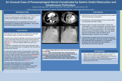
Case Description/Methods: An 80-year-old woman with a history of hiatal hernia presented with intractable nausea, vomiting, and abdominal pain. The patient had a similar presentation a month prior and was noted to have a large hiatal hernia with most of the stomach above the diaphragm on contrast computed tomography (CT) abdomen and pelvis (Figure 1). She was awaiting hiatal hernia repair as an outpatient.
On admission, the patient was found to have leukocytosis, lactic acidosis, and acute kidney injury. A nasogastric tube was placed to decompress the stomach. An upper gastrointestinal series using gastrografin contrast was done to rule out gastric outlet obstruction (GOO). Follow-up films through 90 minutes revealed (GOO) and contrast extravasated at the margin of the hernia and in the left pleural space (Figure 2). Subsequently, CT chest without contrast showed extravasated contrast in the left pleural space consistent with stomach perforation within a large hiatal hernia (Figure 3).
The patient rapidly deteriorated, had increased oxygen requirement, and was placed on a nonrebreather. The patient was evaluated by thoracic surgery, and her family agreed on no surgical intervention due to a poor outcome. She was then transitioned to hospice care.
Discussion:
Hiatal hernias are primarily asymptomatic but may present with life-threatening complications. Complications of paraesophageal hernia are often lethal and associated with high mortality. Elective repair is usually recommended, and this was corroborated in a Finnish retrospective, population-based study. This revealed 16.4% mortality in the conservative group and 2.7% in the surgical group. 13% of the deaths could be avoided with elective surgery, and most deaths were from incarceration. High-risk patients have significantly higher morbidity but not mortality. Unfortunately, our patient could not have this procedure before this complication. Since this condition is associated with potentially lethal complications, it is essential to recognize it early and initiate the right treatment interventions to avert poor outcomes.

Disclosures:
Kwabena O. Asafo-Agyei, MD1, Dominic Amakye, MD2, Prince Djan, MD3, Peyman Roohani, MD1. P1412 - An Unusual Case of Paraesophageal Hernia Complicated by Gastric Outlet Obstruction and Intrathoracic Perforation, ACG 2023 Annual Scientific Meeting Abstracts. Vancouver, BC, Canada: American College of Gastroenterology.
1Christus Highland Hospital, Shreveport, LA; 2Piedmont Athens Regional Medical Center, Athens, GA; 3SOVAH Health, Martinsville, VA
Introduction: Hiatal Hernia occurs with the prolapse of a stomach segment through the diaphragmatic esophageal hiatus. This is common with increases in the incidence with age.
Paraesophageal hernia is associated with a 5% risk of strangulation or perforation with incarceration. This is often lethal without surgical intervention.
We present a case of a large hiatal hernia with acute gastric outlet obstruction and intrathoracic perforation.
Case Description/Methods: An 80-year-old woman with a history of hiatal hernia presented with intractable nausea, vomiting, and abdominal pain. The patient had a similar presentation a month prior and was noted to have a large hiatal hernia with most of the stomach above the diaphragm on contrast computed tomography (CT) abdomen and pelvis (Figure 1). She was awaiting hiatal hernia repair as an outpatient.
On admission, the patient was found to have leukocytosis, lactic acidosis, and acute kidney injury. A nasogastric tube was placed to decompress the stomach. An upper gastrointestinal series using gastrografin contrast was done to rule out gastric outlet obstruction (GOO). Follow-up films through 90 minutes revealed (GOO) and contrast extravasated at the margin of the hernia and in the left pleural space (Figure 2). Subsequently, CT chest without contrast showed extravasated contrast in the left pleural space consistent with stomach perforation within a large hiatal hernia (Figure 3).
The patient rapidly deteriorated, had increased oxygen requirement, and was placed on a nonrebreather. The patient was evaluated by thoracic surgery, and her family agreed on no surgical intervention due to a poor outcome. She was then transitioned to hospice care.
Discussion:
Hiatal hernias are primarily asymptomatic but may present with life-threatening complications. Complications of paraesophageal hernia are often lethal and associated with high mortality. Elective repair is usually recommended, and this was corroborated in a Finnish retrospective, population-based study. This revealed 16.4% mortality in the conservative group and 2.7% in the surgical group. 13% of the deaths could be avoided with elective surgery, and most deaths were from incarceration. High-risk patients have significantly higher morbidity but not mortality. Unfortunately, our patient could not have this procedure before this complication. Since this condition is associated with potentially lethal complications, it is essential to recognize it early and initiate the right treatment interventions to avert poor outcomes.

Figure: Fig 1 reveals a large hiatal hernia with most of the stomach above the diaphragm.
Fig 2 shows a gastrointestinal study using gastrografin contrast which demonstrates GOO and extravasated contrast at the margin of the hernia and in the left pleural space.
Fig 3 showed extravasated contrast in the left pleural space consistent with stomach perforation within a large hiatal hernia.
Fig 2 shows a gastrointestinal study using gastrografin contrast which demonstrates GOO and extravasated contrast at the margin of the hernia and in the left pleural space.
Fig 3 showed extravasated contrast in the left pleural space consistent with stomach perforation within a large hiatal hernia.
Disclosures:
Kwabena Asafo-Agyei indicated no relevant financial relationships.
Dominic Amakye indicated no relevant financial relationships.
Prince Djan indicated no relevant financial relationships.
Peyman Roohani indicated no relevant financial relationships.
Kwabena O. Asafo-Agyei, MD1, Dominic Amakye, MD2, Prince Djan, MD3, Peyman Roohani, MD1. P1412 - An Unusual Case of Paraesophageal Hernia Complicated by Gastric Outlet Obstruction and Intrathoracic Perforation, ACG 2023 Annual Scientific Meeting Abstracts. Vancouver, BC, Canada: American College of Gastroenterology.

