Sunday Poster Session
Category: Stomach
P1391 - Bronchogenic Cyst: Mimicker of Malignant Lymphadenopathy
Sunday, October 22, 2023
3:30 PM - 7:00 PM PT
Location: Exhibit Hall

Has Audio

Victoria Garland, MD
Lenox Hill Hospital, Northwell Health
New York, New York
Presenting Author(s)
Victoria Garland, MD, Isabella Bergagnini, DO, Michael Ma, MD, Beatriz Caraballo Bordon, MD, Elliot Newman, MD, Evin McCabe, MD
Lenox Hill Hospital, Northwell Health, New York, NY
Introduction: Bronchogenic cysts are rare congenital pulmonary malformation of the foregut, typically located in the mediastinum but can occur elsewhere. Most often they are discovered incidentally; however, they can also cause symptoms of compression, rupture or infection. Their varied appearance can also lead to diagnostic uncertainty. We present a case of a patient undergoing gastric malignancy staging found to have a bronchogenic cyst (BC) mimicking lymphadenopathy.
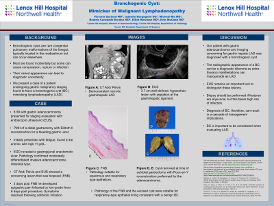
Case Description/Methods: A 61-year-old man newly diagnosed with gastric adenocarcinoma presented for staging evaluation with endoscopic ultrasound (EUS). He had a remote history of distal gastrectomy with Billroth II reconstruction for a bleeding gastric ulcer. He initially presented with fatigue and was found to be anemic with hemoglobin 11 g/dL. He underwent bidirectional endoscopy which revealed ulceration at the gastrojejunal anastomosis. Pathology confirmed moderately differentiated invasive adenocarcinoma, intestinal type. He subsequently underwent a CT scan that demonstrated necrotic gastrohepatic lymphadenopathy (A). EUS revealed a 3.7 cm well-defined and hypoechoic lesion with a septation at the gastrohepatic ligament (B). He underwent fine needle biopsy (FNB). Two days later he developed epigastric pain followed by low-grade fever six days post procedure. Symptoms resolved following initiation of antibiotics.
Pathology from the FNB was notable for squamous and respiratory type epithelium (C, orig. mag. x20). He later underwent a subtotal gastrectomy with Roux-en-Y reconstruction for the adenocarcinoma with removal of the cyst near the lesser curvature (D). Pathology of the excised cyst was again notable for respiratory-type epithelial lining consistent with a benign BC.
Discussion: Our patient with gastric adenocarcinoma and imaging concerning for gastric hepatic lymphadenopathy was diagnosed with a bronchogenic cyst. The radiographic appearance of a BC can be a diagnostic dilemma as extra-thoracic manifestations can masquerade as lymphadenopathy. EUS remains an important tool to distinguish these lesions. Biopsy should be performed if features are equivocal, but this bears high risk of infection. Diagnosis of BC, therefore, can result in a cascade of management implications. Thus, it is important to be considered when evaluating lymphadenopathy.

Disclosures:
Victoria Garland, MD, Isabella Bergagnini, DO, Michael Ma, MD, Beatriz Caraballo Bordon, MD, Elliot Newman, MD, Evin McCabe, MD. P1391 - Bronchogenic Cyst: Mimicker of Malignant Lymphadenopathy, ACG 2023 Annual Scientific Meeting Abstracts. Vancouver, BC, Canada: American College of Gastroenterology.
Lenox Hill Hospital, Northwell Health, New York, NY
Introduction: Bronchogenic cysts are rare congenital pulmonary malformation of the foregut, typically located in the mediastinum but can occur elsewhere. Most often they are discovered incidentally; however, they can also cause symptoms of compression, rupture or infection. Their varied appearance can also lead to diagnostic uncertainty. We present a case of a patient undergoing gastric malignancy staging found to have a bronchogenic cyst (BC) mimicking lymphadenopathy.
Case Description/Methods: A 61-year-old man newly diagnosed with gastric adenocarcinoma presented for staging evaluation with endoscopic ultrasound (EUS). He had a remote history of distal gastrectomy with Billroth II reconstruction for a bleeding gastric ulcer. He initially presented with fatigue and was found to be anemic with hemoglobin 11 g/dL. He underwent bidirectional endoscopy which revealed ulceration at the gastrojejunal anastomosis. Pathology confirmed moderately differentiated invasive adenocarcinoma, intestinal type. He subsequently underwent a CT scan that demonstrated necrotic gastrohepatic lymphadenopathy (A). EUS revealed a 3.7 cm well-defined and hypoechoic lesion with a septation at the gastrohepatic ligament (B). He underwent fine needle biopsy (FNB). Two days later he developed epigastric pain followed by low-grade fever six days post procedure. Symptoms resolved following initiation of antibiotics.
Pathology from the FNB was notable for squamous and respiratory type epithelium (C, orig. mag. x20). He later underwent a subtotal gastrectomy with Roux-en-Y reconstruction for the adenocarcinoma with removal of the cyst near the lesser curvature (D). Pathology of the excised cyst was again notable for respiratory-type epithelial lining consistent with a benign BC.
Discussion: Our patient with gastric adenocarcinoma and imaging concerning for gastric hepatic lymphadenopathy was diagnosed with a bronchogenic cyst. The radiographic appearance of a BC can be a diagnostic dilemma as extra-thoracic manifestations can masquerade as lymphadenopathy. EUS remains an important tool to distinguish these lesions. Biopsy should be performed if features are equivocal, but this bears high risk of infection. Diagnosis of BC, therefore, can result in a cascade of management implications. Thus, it is important to be considered when evaluating lymphadenopathy.

Figure: A) CT scan showing necrotic gastrohepatic lymphadenopathy
B) EUS showing a 3.7 cm well-defined and hypoechoic lesion with a septation at the gastrohepatic ligament
C) Pathology from fine needle biopsy showing squamous and respiratory type epithelium
D) Excised cyst
B) EUS showing a 3.7 cm well-defined and hypoechoic lesion with a septation at the gastrohepatic ligament
C) Pathology from fine needle biopsy showing squamous and respiratory type epithelium
D) Excised cyst
Disclosures:
Victoria Garland indicated no relevant financial relationships.
Isabella Bergagnini indicated no relevant financial relationships.
Michael Ma indicated no relevant financial relationships.
Beatriz Caraballo Bordon indicated no relevant financial relationships.
Elliot Newman indicated no relevant financial relationships.
Evin McCabe indicated no relevant financial relationships.
Victoria Garland, MD, Isabella Bergagnini, DO, Michael Ma, MD, Beatriz Caraballo Bordon, MD, Elliot Newman, MD, Evin McCabe, MD. P1391 - Bronchogenic Cyst: Mimicker of Malignant Lymphadenopathy, ACG 2023 Annual Scientific Meeting Abstracts. Vancouver, BC, Canada: American College of Gastroenterology.
