Sunday Poster Session
Category: Liver
P0960 - Dielectric Properties as a Biomarker for the Diagnosis of Cholestatic Liver Disease: Feasibility Study in Mice Model
Sunday, October 22, 2023
3:30 PM - 7:00 PM PT
Location: Exhibit Hall

Has Audio
- KG
Keerthy Gopalakrishnan, MBBS
The Wright Center for Community Health
Scranton, PA
Presenting Author(s)
Keerthy Gopalakrishnan, MBBS1, Poulami Samaddar, PhD2, Abid Anwar, BS3, Usman Yaqoob, BS2, Nidhi Sakrikar, PhD2, Shivaram Poigai Arunachalam, PhD, DBA2
1The Wright Center for Community Health, Scranton, PA; 2Mayo Clinic, Rochester, MN; 3Mayo Clinic, Naperville, IL
Introduction: Cholestasis is a pathological consequence due to various intrahepatic or extrahepatic defects leading to disruption in bile acid formation, secretion, and excretion. Hepatobiliary conditions due to cholestasis is called cholestatic liver disease characterized by inflammation and tissue fibrosis. It can affect any age group and diagnosis is crucial as some adults maybe asymptomatic and found only during routine lab works. Various biomarkers including ALP, GGT, bile acids and bilirubin are used for diagnosis, but these tests are unreliable with poor sensitivity and specificity. Imaging methods like MRI, CT, ERCP and ultrasound can be helpful only in diagnosing extrahepatic conditions. Diagnosing cholestatic liver disease is still challenging and there is a clear need for novel biomarkers. Microwave diagnostic techniques using dielectric properties have gained popularity in recent years. The purpose of this work was to test the feasibility of using dielectric properties to distinguish mice model with cholestatic liver disease compared to healthy mice.
Methods: Livers from mice fed with either chow or 0.1% DDC (3,5-Diethoxycarbonyl-1,4-Dihydrocollidine) diet for 2 weeks to induce cholestasis were used to diagnose the dielectric properties. The whole mouse liver was tested using an open-ended coaxial slim form probe and a vector network analyzer (VNA, Keysight inc.) within 2-5 minutes after the mouse was sacrificed. The data set was compiled by measuring the ex-vivo dielectric properties of 6 normal and 4 Cholestatic mouse liver tissues in 501 data points per measurement sweep over a frequency range of 0.5 GHz – 20 GHz. Measurements at 7 different areas of the liver were taken at room temperature (21⁰ C).
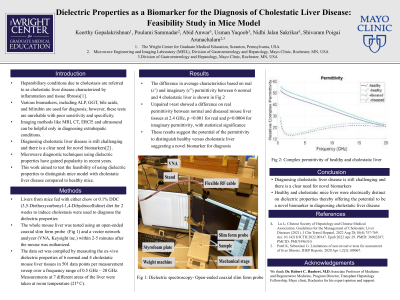
Results: The difference in average characteristics based on real (ɛ’) and imaginary(ɛ”) permittivity between 6 normal and 4 cholestatic liver is shown in Fig 1. Unpaired t-test showed a difference on real permittivity between normal and diseased mouse liver tissues at 2.4 GHz, p < 0.001 for real and p=0.0004 for imaginary permittivity, with statistical significance. These results suggest the potential of the permittivity to distinguish healthy versus cholestatic liver suggesting a novel biomarker for diagnosis.
Discussion: Healthy and cholestatic mice liver were electrically distinct on dielectric properties thereby offers potential to be a novel biomarker in diagnosing cholestatic liver disease.

Disclosures:
Keerthy Gopalakrishnan, MBBS1, Poulami Samaddar, PhD2, Abid Anwar, BS3, Usman Yaqoob, BS2, Nidhi Sakrikar, PhD2, Shivaram Poigai Arunachalam, PhD, DBA2. P0960 - Dielectric Properties as a Biomarker for the Diagnosis of Cholestatic Liver Disease: Feasibility Study in Mice Model, ACG 2023 Annual Scientific Meeting Abstracts. Vancouver, BC, Canada: American College of Gastroenterology.
1The Wright Center for Community Health, Scranton, PA; 2Mayo Clinic, Rochester, MN; 3Mayo Clinic, Naperville, IL
Introduction: Cholestasis is a pathological consequence due to various intrahepatic or extrahepatic defects leading to disruption in bile acid formation, secretion, and excretion. Hepatobiliary conditions due to cholestasis is called cholestatic liver disease characterized by inflammation and tissue fibrosis. It can affect any age group and diagnosis is crucial as some adults maybe asymptomatic and found only during routine lab works. Various biomarkers including ALP, GGT, bile acids and bilirubin are used for diagnosis, but these tests are unreliable with poor sensitivity and specificity. Imaging methods like MRI, CT, ERCP and ultrasound can be helpful only in diagnosing extrahepatic conditions. Diagnosing cholestatic liver disease is still challenging and there is a clear need for novel biomarkers. Microwave diagnostic techniques using dielectric properties have gained popularity in recent years. The purpose of this work was to test the feasibility of using dielectric properties to distinguish mice model with cholestatic liver disease compared to healthy mice.
Methods: Livers from mice fed with either chow or 0.1% DDC (3,5-Diethoxycarbonyl-1,4-Dihydrocollidine) diet for 2 weeks to induce cholestasis were used to diagnose the dielectric properties. The whole mouse liver was tested using an open-ended coaxial slim form probe and a vector network analyzer (VNA, Keysight inc.) within 2-5 minutes after the mouse was sacrificed. The data set was compiled by measuring the ex-vivo dielectric properties of 6 normal and 4 Cholestatic mouse liver tissues in 501 data points per measurement sweep over a frequency range of 0.5 GHz – 20 GHz. Measurements at 7 different areas of the liver were taken at room temperature (21⁰ C).
Results: The difference in average characteristics based on real (ɛ’) and imaginary(ɛ”) permittivity between 6 normal and 4 cholestatic liver is shown in Fig 1. Unpaired t-test showed a difference on real permittivity between normal and diseased mouse liver tissues at 2.4 GHz, p < 0.001 for real and p=0.0004 for imaginary permittivity, with statistical significance. These results suggest the potential of the permittivity to distinguish healthy versus cholestatic liver suggesting a novel biomarker for diagnosis.
Discussion: Healthy and cholestatic mice liver were electrically distinct on dielectric properties thereby offers potential to be a novel biomarker in diagnosing cholestatic liver disease.

Figure: Fig 1: Complex permittivity of healthy and cholestatic liver
Disclosures:
Keerthy Gopalakrishnan indicated no relevant financial relationships.
Poulami Samaddar indicated no relevant financial relationships.
Abid Anwar indicated no relevant financial relationships.
Usman Yaqoob indicated no relevant financial relationships.
Nidhi Sakrikar indicated no relevant financial relationships.
Shivaram Poigai Arunachalam indicated no relevant financial relationships.
Keerthy Gopalakrishnan, MBBS1, Poulami Samaddar, PhD2, Abid Anwar, BS3, Usman Yaqoob, BS2, Nidhi Sakrikar, PhD2, Shivaram Poigai Arunachalam, PhD, DBA2. P0960 - Dielectric Properties as a Biomarker for the Diagnosis of Cholestatic Liver Disease: Feasibility Study in Mice Model, ACG 2023 Annual Scientific Meeting Abstracts. Vancouver, BC, Canada: American College of Gastroenterology.
