Sunday Poster Session
Category: Esophagus
P0486 - Dr. Google Told Me It’s a Dermoid Cyst, a Rare Metastasis of Primary Esophageal Cancer to the Anterior Chest Wall: A Case Report
Sunday, October 22, 2023
3:30 PM - 7:00 PM PT
Location: Exhibit Hall

Has Audio

Mesay H. Asfaw, MD
Howard University Hospital
Takoma Park, MD
Presenting Author(s)
Mesay Asfaw, MD1, Cody King, 2, Lyvia Welborn, 2, Hundaol Yadeta, MD3, Angesom Kibreab, MD3, Farshad Aduli, MD4, Nicholas Azinge, MD3
1Howard University Hospital, Takoma Park, MD; 2Howard University, Washington, WA; 3Howard University Hospital, Washington, WA; 4Howard University Hospital, Washington, DC
Introduction: Esophageal Adenocarcinoma, an aggressive cancer, shows an increasing incidence until recent years. Risk factors include male gender, smoking, obesity, diet, reflux disease, Barrett esophagus, and hiatal hernia. Metastasis to lymph nodes, liver, lungs, bones, and adrenal glands is common, while skin metastasis is rare. The pathogenesis and treatment guidelines for skin metastasis are not well-established. Distant metastasis in esophageal carcinoma signifies a poor prognosis, with a 6% 5-year survival rate. Chemotherapy is the standard treatment, but skin metastasis management lacks clear guidelines. In this case, due to the patient's age, comorbidities, poor functional status, and prognosis, palliative care was chosen after shared decision-making.
Case Description/Methods: A 70-year-old Caucasian man with chronic alcoholism presented with a complex gastrointestinal history including esophageal stricture and chronic pancreatitis. He complained of progressive generalized weakness over a month, leading to an inability to stand unaided. Lower back pain and difficulty eating solid food were also noted. Physical examination revealed a 5.8 x 3.8 x 1.2 cm non-tender anterior chest wall nodule, present for a while, self-diagnosed as a dermoid cyst. CT scan showed diffuse esophageal wall thickening, right paratracheal and left hilar masses suggesting adenopathy, as well as liver, bilateral adrenal, and lung metastases. Esophagogastroduodenoscopy revealed a friable red/pink soft tissue mass (0.5 x 0.3 x 0.1 cm) in the lower third of the esophagus, confirmed as esophageal adenocarcinoma by pathology. The chest wall nodule biopsy indicated poorly differentiated carcinoma with perineural invasion. Positive Cytokeratin 7, Epithelial Membrane Antigen, and rare Cytokeratin 20 staining were observed. Palliative care was initiated due to extensive metastatic disease.
Discussion: Esophageal Adenocarcinoma is a particularly aggressive cancer with the potential to metastasize to distant organs, including the skin. Even though skin metastasis rarely originates from esophageal cancers, they can be one of the first clinical symptoms of underlying internal cancers and therefore should still be considered in the differential diagnosis of patients presenting with skin lesions suspicious for malignancy accompanied by the presence of abdominal masses in diagnostic imaging. This way, favoring early diagnosis and treatment which in turn can improve patient outcomes.

Disclosures:
Mesay Asfaw, MD1, Cody King, 2, Lyvia Welborn, 2, Hundaol Yadeta, MD3, Angesom Kibreab, MD3, Farshad Aduli, MD4, Nicholas Azinge, MD3. P0486 - Dr. Google Told Me It’s a Dermoid Cyst, a Rare Metastasis of Primary Esophageal Cancer to the Anterior Chest Wall: A Case Report, ACG 2023 Annual Scientific Meeting Abstracts. Vancouver, BC, Canada: American College of Gastroenterology.
1Howard University Hospital, Takoma Park, MD; 2Howard University, Washington, WA; 3Howard University Hospital, Washington, WA; 4Howard University Hospital, Washington, DC
Introduction: Esophageal Adenocarcinoma, an aggressive cancer, shows an increasing incidence until recent years. Risk factors include male gender, smoking, obesity, diet, reflux disease, Barrett esophagus, and hiatal hernia. Metastasis to lymph nodes, liver, lungs, bones, and adrenal glands is common, while skin metastasis is rare. The pathogenesis and treatment guidelines for skin metastasis are not well-established. Distant metastasis in esophageal carcinoma signifies a poor prognosis, with a 6% 5-year survival rate. Chemotherapy is the standard treatment, but skin metastasis management lacks clear guidelines. In this case, due to the patient's age, comorbidities, poor functional status, and prognosis, palliative care was chosen after shared decision-making.
Case Description/Methods: A 70-year-old Caucasian man with chronic alcoholism presented with a complex gastrointestinal history including esophageal stricture and chronic pancreatitis. He complained of progressive generalized weakness over a month, leading to an inability to stand unaided. Lower back pain and difficulty eating solid food were also noted. Physical examination revealed a 5.8 x 3.8 x 1.2 cm non-tender anterior chest wall nodule, present for a while, self-diagnosed as a dermoid cyst. CT scan showed diffuse esophageal wall thickening, right paratracheal and left hilar masses suggesting adenopathy, as well as liver, bilateral adrenal, and lung metastases. Esophagogastroduodenoscopy revealed a friable red/pink soft tissue mass (0.5 x 0.3 x 0.1 cm) in the lower third of the esophagus, confirmed as esophageal adenocarcinoma by pathology. The chest wall nodule biopsy indicated poorly differentiated carcinoma with perineural invasion. Positive Cytokeratin 7, Epithelial Membrane Antigen, and rare Cytokeratin 20 staining were observed. Palliative care was initiated due to extensive metastatic disease.
Discussion: Esophageal Adenocarcinoma is a particularly aggressive cancer with the potential to metastasize to distant organs, including the skin. Even though skin metastasis rarely originates from esophageal cancers, they can be one of the first clinical symptoms of underlying internal cancers and therefore should still be considered in the differential diagnosis of patients presenting with skin lesions suspicious for malignancy accompanied by the presence of abdominal masses in diagnostic imaging. This way, favoring early diagnosis and treatment which in turn can improve patient outcomes.

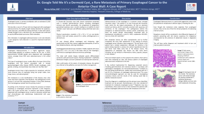
Figure: Images show A) the cutaneous metastasis to the anterior chest wall B) the endoscopic picture of the esophageal mass and C) Histological image of esophageal mass with hematoxylin-eosin stain
Disclosures:
Mesay Asfaw indicated no relevant financial relationships.
Cody King indicated no relevant financial relationships.
Lyvia Welborn indicated no relevant financial relationships.
Hundaol Yadeta indicated no relevant financial relationships.
Angesom Kibreab indicated no relevant financial relationships.
Farshad Aduli indicated no relevant financial relationships.
Nicholas Azinge indicated no relevant financial relationships.
Mesay Asfaw, MD1, Cody King, 2, Lyvia Welborn, 2, Hundaol Yadeta, MD3, Angesom Kibreab, MD3, Farshad Aduli, MD4, Nicholas Azinge, MD3. P0486 - Dr. Google Told Me It’s a Dermoid Cyst, a Rare Metastasis of Primary Esophageal Cancer to the Anterior Chest Wall: A Case Report, ACG 2023 Annual Scientific Meeting Abstracts. Vancouver, BC, Canada: American College of Gastroenterology.
