Sunday Poster Session
Category: Esophagus
P0487 - Double Trouble – A Rare Case of Achalasia Co-Presenting With a Large Epiphrenic Esophageal Diverticulum
Sunday, October 22, 2023
3:30 PM - 7:00 PM PT
Location: Exhibit Hall

Has Audio

Nzube Ekpunobi, MD
Detroit Medical Center
Farmington Hills, MI
Presenting Author(s)
Nzube Ekpunobi, MD1, Robert Goo, MD2
1Detroit Medical Center, Farmington Hills, MI; 2Detroit Medical Center, Commerce, MI
Introduction: Large epiphrenic diverticula ( >5 cm) co-presenting with achalasia are extremely rare, with only a few case reports described. While most patients with either condition are typically asymptomatic, when they present together, common symptoms include dysphagia, regurgitation of undigested food, and weight loss. The risk of esophageal cancer presenting as pseudo-achalasia is also significantly increased when both achalasia and esophageal diverticula are present. Here we present a rare case of achalasia co-presenting with a large epiphrenic esophageal diverticulum.
Case Description/Methods: A 59-year-old woman presented to the ED with severe, colicky right flank pain that worsened with eating. However, on review of systems, she reported a chronic aversion to eating due to nausea, positional heartburn, and difficulty swallowing solids. More concerningly, she admitted to over 100 lbs. of weight loss over the past two years. She was eventually admitted for pyelonephritis. However, gastroenterology was consulted to work up her dysphagia. A barium esophagogram revealed a dilated esophagus with an irregular contour, likely due to functional dysmotility or achalasia. Also visualized was a tuberous, distal esophageal diverticulum measuring 7 x 1.3 cm. Follow-up EGD confirmed these findings, and gastroenterology administered esophageal botulinum, started proton pump inhibitors (PPIs) and recommended outpatient manometry. Esophageal biopsies at the GE junction returned benign results. Cardiothoracic surgery recommended outpatient diverticulum repair, and the patient was discharged home.
Discussion: An epiphrenic diverticulum is a false pulsion diverticulum typically resulting from functional esophageal abnormalities such as achalasia, DES, nutcracker esophagus, and other nonspecific esophageal motility disorders. Surgical intervention is often the definitive treatment. Esophageal cancer is the most feared risk in patients with this co-morbid condition. However, before a diagnosis of cancer, the duration of symptoms is usually at least ten years. The presumed mechanism of cancer development is chronic inflammation secondary to persistent esophageal distention, bacterial overgrowth, and associated GERD. The patient highlighted in our case had biopsies negative for malignancy. However, strong consideration should be given to continued esophageal surveillance, given the high-risk association with malignancy and the lack of data on such patients.

Disclosures:
Nzube Ekpunobi, MD1, Robert Goo, MD2. P0487 - Double Trouble – A Rare Case of Achalasia Co-Presenting With a Large Epiphrenic Esophageal Diverticulum, ACG 2023 Annual Scientific Meeting Abstracts. Vancouver, BC, Canada: American College of Gastroenterology.
1Detroit Medical Center, Farmington Hills, MI; 2Detroit Medical Center, Commerce, MI
Introduction: Large epiphrenic diverticula ( >5 cm) co-presenting with achalasia are extremely rare, with only a few case reports described. While most patients with either condition are typically asymptomatic, when they present together, common symptoms include dysphagia, regurgitation of undigested food, and weight loss. The risk of esophageal cancer presenting as pseudo-achalasia is also significantly increased when both achalasia and esophageal diverticula are present. Here we present a rare case of achalasia co-presenting with a large epiphrenic esophageal diverticulum.
Case Description/Methods: A 59-year-old woman presented to the ED with severe, colicky right flank pain that worsened with eating. However, on review of systems, she reported a chronic aversion to eating due to nausea, positional heartburn, and difficulty swallowing solids. More concerningly, she admitted to over 100 lbs. of weight loss over the past two years. She was eventually admitted for pyelonephritis. However, gastroenterology was consulted to work up her dysphagia. A barium esophagogram revealed a dilated esophagus with an irregular contour, likely due to functional dysmotility or achalasia. Also visualized was a tuberous, distal esophageal diverticulum measuring 7 x 1.3 cm. Follow-up EGD confirmed these findings, and gastroenterology administered esophageal botulinum, started proton pump inhibitors (PPIs) and recommended outpatient manometry. Esophageal biopsies at the GE junction returned benign results. Cardiothoracic surgery recommended outpatient diverticulum repair, and the patient was discharged home.
Discussion: An epiphrenic diverticulum is a false pulsion diverticulum typically resulting from functional esophageal abnormalities such as achalasia, DES, nutcracker esophagus, and other nonspecific esophageal motility disorders. Surgical intervention is often the definitive treatment. Esophageal cancer is the most feared risk in patients with this co-morbid condition. However, before a diagnosis of cancer, the duration of symptoms is usually at least ten years. The presumed mechanism of cancer development is chronic inflammation secondary to persistent esophageal distention, bacterial overgrowth, and associated GERD. The patient highlighted in our case had biopsies negative for malignancy. However, strong consideration should be given to continued esophageal surveillance, given the high-risk association with malignancy and the lack of data on such patients.

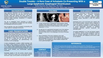
Figure: A. A single-contrast barium esophagogram demonstrating a dilated esophagus with irregular distal contour ("bird's beak"), and a large diverticulum arising from the distal esophagus on the right side measuring 7 x 1.3 cm.
B. An axial view of a computerized tomography scan of the abdomen demonstrating a moderate to large hiatal hernia with moderate amounts of fluid and air in the esophagus to the level of the great vessels.
C. Direct visualization of the lower esophagus using upper endoscopy, revealing a narrowed distal esophagus near the gastroesophageal (GE) junction, with an adjacent large epiphrenic diverticulum and white exudates consistent with chronic gastroesophageal reflux.
Disclosures:
Nzube Ekpunobi indicated no relevant financial relationships.
Robert Goo indicated no relevant financial relationships.
Nzube Ekpunobi, MD1, Robert Goo, MD2. P0487 - Double Trouble – A Rare Case of Achalasia Co-Presenting With a Large Epiphrenic Esophageal Diverticulum, ACG 2023 Annual Scientific Meeting Abstracts. Vancouver, BC, Canada: American College of Gastroenterology.
