Sunday Poster Session
Category: Esophagus
P0491 - Esophageal Carcinoma: An Unlikely Site of Spread
Sunday, October 22, 2023
3:30 PM - 7:00 PM PT
Location: Exhibit Hall

Has Audio

Robert Molina, MPH
University of Texas Medical Branch, John Sealy School of Medicine
Galveston, TX
Presenting Author(s)
Robert Molina, MPH1, Rawan Dayah, MD2, Sreeram Parupudi, MD3, Yasir Ali, MD3
1University of Texas Medical Branch, John Sealy School of Medicine, Galveston, TX; 2University of Texas Medical Branch-Galveston, Galveston, TX; 3University of Texas Medical Branch, Galveston, TX
Introduction: Esophageal carcinomas are one of the most fatal cancers worldwide. These cancers often metastasize to common locations, but in rare occasions an occurrence of this primary tumor can arise in areas not often seen in medicine.
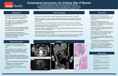
Case Description/Methods: A 70-year-old man presented to an outside hospital for hypovolemic shock. The patient described 2 weeks of weakness, nausea, vomiting, hematuria, and abdominal pain localized to the epigastric and left upper quadrant. His past medical history was significant for squamous cell carcinoma of the esophagus, hypertension, and seizures. Initial studies revealed conjugated hyperbilirubinemia with transaminitis. Evaluation included an abdominal and pelvic CT scan revealing a pancreatic head mass, distal pancreatic parenchymal atrophy, significant intrahepatic and extrahepatic bile duct dilation, and several enlarged retroperitoneal lymph nodes. He was transferred to our hospital for further evaluation. Physical exam was notable for mild tenderness to palpation in the RUQ and LLQ and decreased strength in the right upper extremity. Relevant laboratory data included a conjugated bilirubin 10.5 mg/dL; alkaline phosphatase 764 U/L; ALT 197 U/L; AST 170 U/L; CA 19-9 of 518.9 U/mL. Upper endoscopic ultrasound revealed a mass localized to the pancreatic head and enlarged lymph nodes in the peripancreatic region. Fine needle aspiration of the mass revealed metastatic SCC, consistent with his prior esophageal SCC.
Discussion: This patient’s history of metastatic esophageal cancer, and the occurrence of a new pancreatic lesion, led to the diagnosis of a rare phenomenon of a primary esophageal SCC metastasizing to the pancreatic head. Malignancies of the esophagus include squamous cell carcinoma and adenocarcinoma, with adenocarcinoma being more common in North America. Esophageal SCC has often metastasized by the time of diagnosis, with lymph nodes, liver, lung, bone, and brain being the most common sites. Metastasis to the pancreas is quite rare, and only a few cases with metastases occurring long after initial diagnosis have been reported. The occurrence of pancreatic metastasis from esophageal squamous cell carcinoma is reported around 2.9% . Esophageal carcinoma carries a poor prognosis with only 15%-25% of patients surviving for 5 years post diagnosis. Our patient’s rare presentation highlights the unpredictable nature of this malignancy and the importance of reducing risk factors that predispose to the development of these deadly tumors.

Disclosures:
Robert Molina, MPH1, Rawan Dayah, MD2, Sreeram Parupudi, MD3, Yasir Ali, MD3. P0491 - Esophageal Carcinoma: An Unlikely Site of Spread, ACG 2023 Annual Scientific Meeting Abstracts. Vancouver, BC, Canada: American College of Gastroenterology.
1University of Texas Medical Branch, John Sealy School of Medicine, Galveston, TX; 2University of Texas Medical Branch-Galveston, Galveston, TX; 3University of Texas Medical Branch, Galveston, TX
Introduction: Esophageal carcinomas are one of the most fatal cancers worldwide. These cancers often metastasize to common locations, but in rare occasions an occurrence of this primary tumor can arise in areas not often seen in medicine.
Case Description/Methods: A 70-year-old man presented to an outside hospital for hypovolemic shock. The patient described 2 weeks of weakness, nausea, vomiting, hematuria, and abdominal pain localized to the epigastric and left upper quadrant. His past medical history was significant for squamous cell carcinoma of the esophagus, hypertension, and seizures. Initial studies revealed conjugated hyperbilirubinemia with transaminitis. Evaluation included an abdominal and pelvic CT scan revealing a pancreatic head mass, distal pancreatic parenchymal atrophy, significant intrahepatic and extrahepatic bile duct dilation, and several enlarged retroperitoneal lymph nodes. He was transferred to our hospital for further evaluation. Physical exam was notable for mild tenderness to palpation in the RUQ and LLQ and decreased strength in the right upper extremity. Relevant laboratory data included a conjugated bilirubin 10.5 mg/dL; alkaline phosphatase 764 U/L; ALT 197 U/L; AST 170 U/L; CA 19-9 of 518.9 U/mL. Upper endoscopic ultrasound revealed a mass localized to the pancreatic head and enlarged lymph nodes in the peripancreatic region. Fine needle aspiration of the mass revealed metastatic SCC, consistent with his prior esophageal SCC.
Discussion: This patient’s history of metastatic esophageal cancer, and the occurrence of a new pancreatic lesion, led to the diagnosis of a rare phenomenon of a primary esophageal SCC metastasizing to the pancreatic head. Malignancies of the esophagus include squamous cell carcinoma and adenocarcinoma, with adenocarcinoma being more common in North America. Esophageal SCC has often metastasized by the time of diagnosis, with lymph nodes, liver, lung, bone, and brain being the most common sites. Metastasis to the pancreas is quite rare, and only a few cases with metastases occurring long after initial diagnosis have been reported. The occurrence of pancreatic metastasis from esophageal squamous cell carcinoma is reported around 2.9% . Esophageal carcinoma carries a poor prognosis with only 15%-25% of patients surviving for 5 years post diagnosis. Our patient’s rare presentation highlights the unpredictable nature of this malignancy and the importance of reducing risk factors that predispose to the development of these deadly tumors.

Figure: Figure 1: CTAP displaying mass located in the pancreatic head
Figure 2: Endoscopic ultrasound revealing irregular pancreatic head mass
Figure 3: CTAP displaying pancreatic head mass with central necrosis
Figure 4 & 5: Pathology displaying malignant cells in background of necrosis
Figure 2: Endoscopic ultrasound revealing irregular pancreatic head mass
Figure 3: CTAP displaying pancreatic head mass with central necrosis
Figure 4 & 5: Pathology displaying malignant cells in background of necrosis
Disclosures:
Robert Molina indicated no relevant financial relationships.
Rawan Dayah indicated no relevant financial relationships.
Sreeram Parupudi indicated no relevant financial relationships.
Yasir Ali indicated no relevant financial relationships.
Robert Molina, MPH1, Rawan Dayah, MD2, Sreeram Parupudi, MD3, Yasir Ali, MD3. P0491 - Esophageal Carcinoma: An Unlikely Site of Spread, ACG 2023 Annual Scientific Meeting Abstracts. Vancouver, BC, Canada: American College of Gastroenterology.
