Sunday Poster Session
Category: Esophagus
P0498 - Gastric Polyp Prolapsing into the Esophagus: A Curious Case of Dysphagia
Sunday, October 22, 2023
3:30 PM - 7:00 PM PT
Location: Exhibit Hall

Has Audio

Tanisha Kalra, MD
SUNY Downstate Health Sciences University/NYCHHC Kings County Hospital
New York, NY
Presenting Author(s)
Tanisha Kalra, MD1, Nihaal P.. Karnik, MD2, Savanna Thor, DO, MPH3
1SUNY Downstate Health Sciences University/NYCHHC Kings County Hospital, New York, NY; 2SUNY Downstate Health Sciences University, New York, NY; 3SUNY Downstate Health Sciences University, Brooklyn, NY
Introduction: Esophageal dysphagia can occur due to mechanical obstructions (esophageal webs, rings, strictures, malignancies) or motility disorders (esophageal spasm, gastroesophageal reflux disease (GERD), achalasia). We present a unique case of dysphagia due to a hyperplastic gastric cardia polyp prolapsing into the gastro-esophageal (GE) junction causing esophageal obstruction.
Case Description/Methods: 44-year-old woman with Down syndrome, Grave’s disease, GERD, obstructive sleep apnea presented with dysphagia, postprandial choking, and regurgitation of solid food after each meal as noted by her caregiver. GERD was diagnosed two years prior to the presentation and she was taking famotidine 40 mg twice daily(BID).
A complete blood count, liver and kidney function tests, and chest x-ray were normal. Evaluation by speech pathology and modified barium swallow excluded oropharyngeal dysphagia. A CT scan of the abdomen/pelvis demonstrated a lesion adjacent to the GE junction. Esophagogastroduodenoscopy (EGD) showed a 1.5 x 1.2 cm, semi-pedunculated polyp arising from gastric cardia. This polyp was seen prolapsing and ball-valving into the GE junction. Esophageal biopsies excluded eosinophilic esophagitis. But, dysphagia persisted despite starting omeprazole 40 mg BID for GERD.
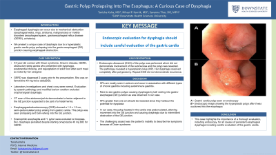
Hence, an endoscopic ultrasound (EUS) of the polyp was performed which did not demonstrate involvement of the submucosa and the polyp was resected. The pathology revealed a hyperplastic polyp (HP) without evidence of Helicobacter Pylori. Her dysphagia resolved completely after polypectomy. Repeat EGD did not demonstrate recurrence.
Discussion: HPs are known to occur in association with different types of chronic gastritis including autoimmune gastritis, however that was not evident here. HPs are mostly seen in the antrum, and cases of gastric outlet obstruction due to HP have been described in the literature, but dysphagia due to HP obstructing the GE junction is rare. In our case, the polyp located in the cardia was pedunculated, allowing movement into the GE junction and causing dysphagia due to intermittent obstruction of the GE junction. HPs greater than one cm should be resected since they harbor potential for dysplasia. Another challenging aspect was the patient’s inability to describe her symptoms because of Down syndrome. This case highlights the importance of a thorough evaluation, including endoscopy, for all causes of persistent esophageal dysphagia.

Disclosures:
Tanisha Kalra, MD1, Nihaal P.. Karnik, MD2, Savanna Thor, DO, MPH3. P0498 - Gastric Polyp Prolapsing into the Esophagus: A Curious Case of Dysphagia, ACG 2023 Annual Scientific Meeting Abstracts. Vancouver, BC, Canada: American College of Gastroenterology.
1SUNY Downstate Health Sciences University/NYCHHC Kings County Hospital, New York, NY; 2SUNY Downstate Health Sciences University, New York, NY; 3SUNY Downstate Health Sciences University, Brooklyn, NY
Introduction: Esophageal dysphagia can occur due to mechanical obstructions (esophageal webs, rings, strictures, malignancies) or motility disorders (esophageal spasm, gastroesophageal reflux disease (GERD), achalasia). We present a unique case of dysphagia due to a hyperplastic gastric cardia polyp prolapsing into the gastro-esophageal (GE) junction causing esophageal obstruction.
Case Description/Methods: 44-year-old woman with Down syndrome, Grave’s disease, GERD, obstructive sleep apnea presented with dysphagia, postprandial choking, and regurgitation of solid food after each meal as noted by her caregiver. GERD was diagnosed two years prior to the presentation and she was taking famotidine 40 mg twice daily(BID).
A complete blood count, liver and kidney function tests, and chest x-ray were normal. Evaluation by speech pathology and modified barium swallow excluded oropharyngeal dysphagia. A CT scan of the abdomen/pelvis demonstrated a lesion adjacent to the GE junction. Esophagogastroduodenoscopy (EGD) showed a 1.5 x 1.2 cm, semi-pedunculated polyp arising from gastric cardia. This polyp was seen prolapsing and ball-valving into the GE junction. Esophageal biopsies excluded eosinophilic esophagitis. But, dysphagia persisted despite starting omeprazole 40 mg BID for GERD.
Hence, an endoscopic ultrasound (EUS) of the polyp was performed which did not demonstrate involvement of the submucosa and the polyp was resected. The pathology revealed a hyperplastic polyp (HP) without evidence of Helicobacter Pylori. Her dysphagia resolved completely after polypectomy. Repeat EGD did not demonstrate recurrence.
Discussion: HPs are known to occur in association with different types of chronic gastritis including autoimmune gastritis, however that was not evident here. HPs are mostly seen in the antrum, and cases of gastric outlet obstruction due to HP have been described in the literature, but dysphagia due to HP obstructing the GE junction is rare. In our case, the polyp located in the cardia was pedunculated, allowing movement into the GE junction and causing dysphagia due to intermittent obstruction of the GE junction. HPs greater than one cm should be resected since they harbor potential for dysplasia. Another challenging aspect was the patient’s inability to describe her symptoms because of Down syndrome. This case highlights the importance of a thorough evaluation, including endoscopy, for all causes of persistent esophageal dysphagia.

Figure: A - Gastric cardia polyp seen on endoscopy
B - Endoscopic image showing the hyperplastic polyp after it was suctioned into the esophagus
B - Endoscopic image showing the hyperplastic polyp after it was suctioned into the esophagus
Disclosures:
Tanisha Kalra indicated no relevant financial relationships.
Nihaal Karnik indicated no relevant financial relationships.
Savanna Thor indicated no relevant financial relationships.
Tanisha Kalra, MD1, Nihaal P.. Karnik, MD2, Savanna Thor, DO, MPH3. P0498 - Gastric Polyp Prolapsing into the Esophagus: A Curious Case of Dysphagia, ACG 2023 Annual Scientific Meeting Abstracts. Vancouver, BC, Canada: American College of Gastroenterology.
