Sunday Poster Session
Category: Colon
P0267 - A Unique Presentation of Familial Idiopathic Colonic Varices
Sunday, October 22, 2023
3:30 PM - 7:00 PM PT
Location: Exhibit Hall

Has Audio

John Paul Gallagher, MD
University of Nebraska Medical Center
Omaha, NE
Presenting Author(s)
Award: Presidential Poster Award
John Gallagher, MD, Bill Quach, MD, Tomoki Sempokuya, MD, Anita Sivaraman, MD
University of Nebraska Medical Center, Omaha, NE
Introduction: Colonic varices typically occur in the setting of portal hypertension and patients may present with rectal bleeding or occult anemia. Idiopathic colonic varices in the absence of cirrhosis occur infrequently and can involve the entire colon. We present a case of a 54-year-old female who underwent diagnostic colonoscopy and was incidentally found to have colonic varices in the setting of new sigmoid adenocarcinoma. Her 38-year-old daughter was found to have similar varices.
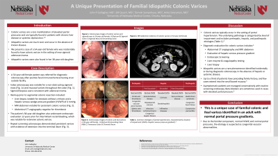
Case Description/Methods: A 54-year-old female patient was referred to our institution for diagnostic colonoscopy after positive fecal immunochemical testing. Index colonoscopy was notable for a non-obstructing sigmoid mass with central depression and serpiginous blue vasculature in the upper rectum extending throughout the colon, without stigmata of bleeding. Biopsies were consistent with adenocarcinoma (Fig. 1). Workup prior to segmental colonic resection included liver biopsy notable for steatosis without cirrhosis and a portal pressure gradient of 4 mmHg.
On additional evaluation, it was discovered that the patient’s 38-year-old daughter previously had intermittent rectal bleeding and underwent endoscopic evaluation 12 years prior. It was notable for colonic varices with ileal extension. Liver biopsy was negative for cirrhosis. Due to her mother’s colon malignancy, she underwent screening colonoscopy which demonstrated similar venous findings (Fig. 2).
Discussion: Idiopathic varices are a rare phenomenon identified incidentally or during diagnostic colonoscopy. Up to a third of patients have preceding family history, and few cases extend into the small bowel mucosa. Prior case reports documented normal portal pressure gradients in individuals with idiopathic varices, but this is the first presentation of familial varices with normal pressure gradients.
Most patients are managed conservatively, and treatment is reserved for recurrent or significant gastrointestinal bleeding. Successful treatments have included partial colectomy, venous coil embolization, balloon-occluded retrograde transvenous obliteration, or angioplasty in cases with vascular involvement. Beta-blockers and somatostatin analogues can be used to reduce portal pressure in patients with elevated portal pressures. There have been insufficient cases to determine if patients with idiopathic varices are at increased risk for malignancy. While there seems to be a hereditary component, specific genes have not yet been identified to identify family members at risk.

Disclosures:
John Gallagher, MD, Bill Quach, MD, Tomoki Sempokuya, MD, Anita Sivaraman, MD. P0267 - A Unique Presentation of Familial Idiopathic Colonic Varices, ACG 2023 Annual Scientific Meeting Abstracts. Vancouver, BC, Canada: American College of Gastroenterology.
John Gallagher, MD, Bill Quach, MD, Tomoki Sempokuya, MD, Anita Sivaraman, MD
University of Nebraska Medical Center, Omaha, NE
Introduction: Colonic varices typically occur in the setting of portal hypertension and patients may present with rectal bleeding or occult anemia. Idiopathic colonic varices in the absence of cirrhosis occur infrequently and can involve the entire colon. We present a case of a 54-year-old female who underwent diagnostic colonoscopy and was incidentally found to have colonic varices in the setting of new sigmoid adenocarcinoma. Her 38-year-old daughter was found to have similar varices.
Case Description/Methods: A 54-year-old female patient was referred to our institution for diagnostic colonoscopy after positive fecal immunochemical testing. Index colonoscopy was notable for a non-obstructing sigmoid mass with central depression and serpiginous blue vasculature in the upper rectum extending throughout the colon, without stigmata of bleeding. Biopsies were consistent with adenocarcinoma (Fig. 1). Workup prior to segmental colonic resection included liver biopsy notable for steatosis without cirrhosis and a portal pressure gradient of 4 mmHg.
On additional evaluation, it was discovered that the patient’s 38-year-old daughter previously had intermittent rectal bleeding and underwent endoscopic evaluation 12 years prior. It was notable for colonic varices with ileal extension. Liver biopsy was negative for cirrhosis. Due to her mother’s colon malignancy, she underwent screening colonoscopy which demonstrated similar venous findings (Fig. 2).
Discussion: Idiopathic varices are a rare phenomenon identified incidentally or during diagnostic colonoscopy. Up to a third of patients have preceding family history, and few cases extend into the small bowel mucosa. Prior case reports documented normal portal pressure gradients in individuals with idiopathic varices, but this is the first presentation of familial varices with normal pressure gradients.
Most patients are managed conservatively, and treatment is reserved for recurrent or significant gastrointestinal bleeding. Successful treatments have included partial colectomy, venous coil embolization, balloon-occluded retrograde transvenous obliteration, or angioplasty in cases with vascular involvement. Beta-blockers and somatostatin analogues can be used to reduce portal pressure in patients with elevated portal pressures. There have been insufficient cases to determine if patients with idiopathic varices are at increased risk for malignancy. While there seems to be a hereditary component, specific genes have not yet been identified to identify family members at risk.

Figure: Figure 1. Colonoscopy images of colonic varices and sigmoid mass in 54-year-old female. 1a) Rectum 1b) Sigmoid Colon 1c) Sigmoid Mass 1d) Descending Colon. Figure 2. Colonoscopy images of colonic and ileal varices in 38-year-old female.
2a) Rectum 2b) Ascending Colon (NBI) 2c) Transverse Colon 2d) Terminal Ileum
2a) Rectum 2b) Ascending Colon (NBI) 2c) Transverse Colon 2d) Terminal Ileum
Disclosures:
John Gallagher indicated no relevant financial relationships.
Bill Quach indicated no relevant financial relationships.
Tomoki Sempokuya indicated no relevant financial relationships.
Anita Sivaraman indicated no relevant financial relationships.
John Gallagher, MD, Bill Quach, MD, Tomoki Sempokuya, MD, Anita Sivaraman, MD. P0267 - A Unique Presentation of Familial Idiopathic Colonic Varices, ACG 2023 Annual Scientific Meeting Abstracts. Vancouver, BC, Canada: American College of Gastroenterology.

