Sunday Poster Session
Category: Colon
P0276 - Cecal Bascule: A Complication of Cardiac Disease?
Sunday, October 22, 2023
3:30 PM - 7:00 PM PT
Location: Exhibit Hall

Has Audio

Maria F. Winte, MD
Thomas Jefferson University Hospital
Philadelphia, PA
Presenting Author(s)
Maria F. Winte, MD1, Fred Karaisz, MD2, Naman Upadhyay, MD3, Raina Shivashankar, MD, FACG1, Indranee Rajapreyar, MD1, Caitlyn Costanzo, MD1
1Thomas Jefferson University Hospital, Philadelphia, PA; 2The Ohio State University Wexner Medical Center, Columbus, OH; 3Cooper University Health Care, Camden, NJ
Introduction: Cecal bascule is a rare type of colonic volvulus. It is believed that its pathogenesis involves a congenital predisposition and a physiologic stressor, such as ischemia. Consequently, it is a source of morbidity and mortality among patients with cardiac diseases.
Case Description/Methods: A 55-year-old man with ischemic cardiomyopathy presented to the hospital with cardiogenic shock. He received an intra-aortic balloon pump for hemodynamic support while awaiting heart transplant evaluation. His hospital course was complicated by intra-aortic balloon pump thrombosis and stroke. On hospital day 170, he developed new-onset abdominal pain and distension. CT of the abdomen showed that the cecum had shifted toward the midline. Serial abdominal X-rays showed worsening colonic dilation and repeat CT confirmed a mobile cecal bascule. After a multi-disciplinary discussion, he underwent open ileocectomy on hospital day 177. His post-operative course was uneventful, and he underwent heart transplant on hospital day 249.
Discussion: Cecal volvulus is classified into three different types: Type 1 develops from clockwise axial torsion of the cecum, Type 2 arises from torsion of the cecum and terminal ileum that is usually in the counterclockwise rotation, and Type 3, known as a cecal bascule, results from upward angulation of the cecum. The incidence of cecal volvulus is increasing yearly and continues to be associated with high mortality rates. It is believed that a predisposition to cecal volvuli requires mobility of the ascending colon, which can be congenital or acquired, and occurs in up to 25% of the population.
Multiple case reports of cecal volvulus in patients with recent cardiothoracic surgery, cardiomyopathy, or coronary artery disease suggest a possible ischemic or cardiac predilection. While the overall incidence of GI complications in cardiac surgery is low (1.1%), mortality due to such complications is high (33%). Likewise, cecal volvulus is associated with a mortality of 10% to 40%. The diagnosis of cecal volvulus involves abdominal radiography and/or cross-sectional imaging. Surgical options for management include cecopexy, cecostomy, colectomy, or manual detorsion; these options must be considered case-by-case, as recurrence rate is high without resection, but resection can carry higher mortality. While successful colonic decompression of cecal bascule has been described, most cases of cecal volvulus are treated with surgical resection.

Disclosures:
Maria F. Winte, MD1, Fred Karaisz, MD2, Naman Upadhyay, MD3, Raina Shivashankar, MD, FACG1, Indranee Rajapreyar, MD1, Caitlyn Costanzo, MD1. P0276 - Cecal Bascule: A Complication of Cardiac Disease?, ACG 2023 Annual Scientific Meeting Abstracts. Vancouver, BC, Canada: American College of Gastroenterology.
1Thomas Jefferson University Hospital, Philadelphia, PA; 2The Ohio State University Wexner Medical Center, Columbus, OH; 3Cooper University Health Care, Camden, NJ
Introduction: Cecal bascule is a rare type of colonic volvulus. It is believed that its pathogenesis involves a congenital predisposition and a physiologic stressor, such as ischemia. Consequently, it is a source of morbidity and mortality among patients with cardiac diseases.
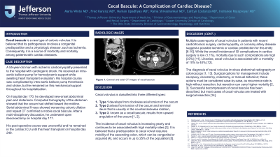
Case Description/Methods: A 55-year-old man with ischemic cardiomyopathy presented to the hospital with cardiogenic shock. He received an intra-aortic balloon pump for hemodynamic support while awaiting heart transplant evaluation. His hospital course was complicated by intra-aortic balloon pump thrombosis and stroke. On hospital day 170, he developed new-onset abdominal pain and distension. CT of the abdomen showed that the cecum had shifted toward the midline. Serial abdominal X-rays showed worsening colonic dilation and repeat CT confirmed a mobile cecal bascule. After a multi-disciplinary discussion, he underwent open ileocectomy on hospital day 177. His post-operative course was uneventful, and he underwent heart transplant on hospital day 249.
Discussion: Cecal volvulus is classified into three different types: Type 1 develops from clockwise axial torsion of the cecum, Type 2 arises from torsion of the cecum and terminal ileum that is usually in the counterclockwise rotation, and Type 3, known as a cecal bascule, results from upward angulation of the cecum. The incidence of cecal volvulus is increasing yearly and continues to be associated with high mortality rates. It is believed that a predisposition to cecal volvuli requires mobility of the ascending colon, which can be congenital or acquired, and occurs in up to 25% of the population.
Multiple case reports of cecal volvulus in patients with recent cardiothoracic surgery, cardiomyopathy, or coronary artery disease suggest a possible ischemic or cardiac predilection. While the overall incidence of GI complications in cardiac surgery is low (1.1%), mortality due to such complications is high (33%). Likewise, cecal volvulus is associated with a mortality of 10% to 40%. The diagnosis of cecal volvulus involves abdominal radiography and/or cross-sectional imaging. Surgical options for management include cecopexy, cecostomy, colectomy, or manual detorsion; these options must be considered case-by-case, as recurrence rate is high without resection, but resection can carry higher mortality. While successful colonic decompression of cecal bascule has been described, most cases of cecal volvulus are treated with surgical resection.

Figure: Figure 1. Coronal CT image of cecal bascule.
Disclosures:
Maria Winte indicated no relevant financial relationships.
Fred Karaisz indicated no relevant financial relationships.
Naman Upadhyay indicated no relevant financial relationships.
Raina Shivashankar: Abbvie – Grant/Research Support, Speakers Bureau. Bristol Meyers Squibb – Speakers Bureau. Janssen – Advisory Committee/Board Member, Grant/Research Support.
Indranee Rajapreyar indicated no relevant financial relationships.
Caitlyn Costanzo indicated no relevant financial relationships.
Maria F. Winte, MD1, Fred Karaisz, MD2, Naman Upadhyay, MD3, Raina Shivashankar, MD, FACG1, Indranee Rajapreyar, MD1, Caitlyn Costanzo, MD1. P0276 - Cecal Bascule: A Complication of Cardiac Disease?, ACG 2023 Annual Scientific Meeting Abstracts. Vancouver, BC, Canada: American College of Gastroenterology.
