Sunday Poster Session
Category: Colon
P0278 - The Scope of Telescoping, Seen on Endoscope
Sunday, October 22, 2023
3:30 PM - 7:00 PM PT
Location: Exhibit Hall

Has Audio

Tevan Y. Luong, BS
UC Irvine School of Medicine
Tustin, CA
Presenting Author(s)
Tevan Y.. Luong, BS1, C Gregory Albers, MD, FACG2, Natalie Hall, MD3, Valery Vilchez, MD4, Steve Huy D. Phan, BS, BA4, Whitney Li, BA5, Rishi Vermani, BS6
1UC Irvine School of Medicine, Tustin, CA; 2UC Irvine Digestive Health Institute, Newport Coast, CA; 3UC Irvine Digestive Health Institute, Orange, CA; 4UC Irvine Digestive Health Institute, Irvine, CA; 5UC Irvine School of Medicine, San Jose, CA; 6UC Irvine School of Medicine, Irvine, CA
Introduction: Gastrointestinal intussusception (GII) is a condition characterized by the telescoping of bowel tissue, 90% of which in adults is caused by masses within the bowel. Intussusception is rare in adults, accounting for less than 5% of cases. GIIs primarily occur at the ileo-colic junction, but may rarely present exclusively in the colon in approximately 5-10% of cases. In this case, we present a unique instance of rectosigmoid intussusception (RSI) secondary to colorectal cancer in an adult male. Understanding the characteristics of intussusception, particularly in the rectosigmoid junction, may contribute to the development of specific diagnostic, management, and treatment strategies, potentially leading to improved patient outcomes.
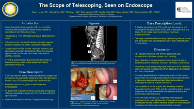
Case Description/Methods: A 51-year-old man with a history of abdominal surgery was admitted to the hospital due to abdominal pain, hematochezia, anemia, and a 50-pound weight loss over the past 6 months. Abdominal exam at presentation and throughout hospital stay was benign. An abdominal contrast-enhanced computed tomography (CT) scan revealed evidence of a mass within the sigmoid colon. Due to improved symptoms and history of negative colonoscopy, a flexible sigmoidoscopy (FS) confirmed the presence of a RSI with a lead point due to a friable 10cm mass, confirmed on pathology to be a mucinous adenocarcinoma. Laparoscopic resection of the mass was performed, and the patient was discharged two days later without further complications.
Discussion: RSI typically manifests with chronic abdominal pain, hematochezia, diarrhea, nausea, and vomiting. Historically, laparoscopy diagnoses the cause of GII. However, choice of surgical management of GII depends on the location and size of the lesion. Early detection of intussusception is vital, given the risk of developing bowel ischemia, necrosis, perforation, and sepsis. This case presented with a fascinating twist—a 10cm mass detected by FS, which paradoxically presented with resolution of abdominal pain and intermittent diarrheal symptoms following the patient’s hospital admission. The utilization of FS as a quick and minimally invasive diagnostic tool to verify RSIs and the underlying lesion equips physicians with vital information for treatment planning and accelerates patient recovery. Thus, this case serves as a compelling teaching point, emphasizing the potential benefits of integrating endoscopic procedures into the current standard of care for assessing and managing rare obstructive gastrointestinal disorders.

Disclosures:
Tevan Y.. Luong, BS1, C Gregory Albers, MD, FACG2, Natalie Hall, MD3, Valery Vilchez, MD4, Steve Huy D. Phan, BS, BA4, Whitney Li, BA5, Rishi Vermani, BS6. P0278 - The Scope of Telescoping, Seen on Endoscope, ACG 2023 Annual Scientific Meeting Abstracts. Vancouver, BC, Canada: American College of Gastroenterology.
1UC Irvine School of Medicine, Tustin, CA; 2UC Irvine Digestive Health Institute, Newport Coast, CA; 3UC Irvine Digestive Health Institute, Orange, CA; 4UC Irvine Digestive Health Institute, Irvine, CA; 5UC Irvine School of Medicine, San Jose, CA; 6UC Irvine School of Medicine, Irvine, CA
Introduction: Gastrointestinal intussusception (GII) is a condition characterized by the telescoping of bowel tissue, 90% of which in adults is caused by masses within the bowel. Intussusception is rare in adults, accounting for less than 5% of cases. GIIs primarily occur at the ileo-colic junction, but may rarely present exclusively in the colon in approximately 5-10% of cases. In this case, we present a unique instance of rectosigmoid intussusception (RSI) secondary to colorectal cancer in an adult male. Understanding the characteristics of intussusception, particularly in the rectosigmoid junction, may contribute to the development of specific diagnostic, management, and treatment strategies, potentially leading to improved patient outcomes.
Case Description/Methods: A 51-year-old man with a history of abdominal surgery was admitted to the hospital due to abdominal pain, hematochezia, anemia, and a 50-pound weight loss over the past 6 months. Abdominal exam at presentation and throughout hospital stay was benign. An abdominal contrast-enhanced computed tomography (CT) scan revealed evidence of a mass within the sigmoid colon. Due to improved symptoms and history of negative colonoscopy, a flexible sigmoidoscopy (FS) confirmed the presence of a RSI with a lead point due to a friable 10cm mass, confirmed on pathology to be a mucinous adenocarcinoma. Laparoscopic resection of the mass was performed, and the patient was discharged two days later without further complications.
Discussion: RSI typically manifests with chronic abdominal pain, hematochezia, diarrhea, nausea, and vomiting. Historically, laparoscopy diagnoses the cause of GII. However, choice of surgical management of GII depends on the location and size of the lesion. Early detection of intussusception is vital, given the risk of developing bowel ischemia, necrosis, perforation, and sepsis. This case presented with a fascinating twist—a 10cm mass detected by FS, which paradoxically presented with resolution of abdominal pain and intermittent diarrheal symptoms following the patient’s hospital admission. The utilization of FS as a quick and minimally invasive diagnostic tool to verify RSIs and the underlying lesion equips physicians with vital information for treatment planning and accelerates patient recovery. Thus, this case serves as a compelling teaching point, emphasizing the potential benefits of integrating endoscopic procedures into the current standard of care for assessing and managing rare obstructive gastrointestinal disorders.

Figure: Figure 1.
A. Surgical excision of rectosigmoid colon
B. Flexible Sigmoidoscopy: Proximal rectosigmoid intussusception
C. Flexible Sigmoidoscopy: Proximal rectosigmoid intussusception with 10cm mucinous adenocarcinoma
A. Surgical excision of rectosigmoid colon
B. Flexible Sigmoidoscopy: Proximal rectosigmoid intussusception
C. Flexible Sigmoidoscopy: Proximal rectosigmoid intussusception with 10cm mucinous adenocarcinoma
Disclosures:
Tevan Luong indicated no relevant financial relationships.
C Gregory Albers: Abbvie – Speakers Bureau. DSI Pharmaceuticals – Speakers Bureau. Nestle Health Sciences – Speakers Bureau. Phathom Pharmaceuticals – Speakers Bureau. Salix Pharmaceuticals – Speakers Bureau.
Natalie Hall indicated no relevant financial relationships.
Valery Vilchez indicated no relevant financial relationships.
Steve Huy Phan indicated no relevant financial relationships.
Whitney Li indicated no relevant financial relationships.
Rishi Vermani indicated no relevant financial relationships.
Tevan Y.. Luong, BS1, C Gregory Albers, MD, FACG2, Natalie Hall, MD3, Valery Vilchez, MD4, Steve Huy D. Phan, BS, BA4, Whitney Li, BA5, Rishi Vermani, BS6. P0278 - The Scope of Telescoping, Seen on Endoscope, ACG 2023 Annual Scientific Meeting Abstracts. Vancouver, BC, Canada: American College of Gastroenterology.
