Sunday Poster Session
Category: Colon
P0282 - Ischemic Colitis in Williams Syndrome: A Case of a Rare Complication of a Rare Disease
Sunday, October 22, 2023
3:30 PM - 7:00 PM PT
Location: Exhibit Hall

Has Audio
- DA
Daniel J. Alvarez, MD
Montefiore Medical Center/Albert Einstein College of Medicine
Bronx, NY
Presenting Author(s)
Daniel J. Alvarez, MD, Alexander Abadir, MD, Harvey Rosenberg, MD
Montefiore Medical Center/Albert Einstein College of Medicine, Bronx, NY
Introduction: Williams syndrome, first described in 1961 by J.C.P Williams, is an autosomal dominant disease caused by a hemizygous deletion on chromosome 7q11.23. This syndrome, with an estimated prevalence of 1:7,500 to 1:20,000, is characterized by distinctive facies, developmental delay, connective tissue abnormalities and involves multiple systems including cardiovascular, endocrine and gastrointestinal. Commonly described gastrointestinal features include feeding difficulties in children (70%), constipation (50%), umbilical and inguinal hernias (40/50%), diverticulosis (30%), and rectal prolapse (15%). We report the rare case of ischemic colitis in a middle-aged adult with Williams syndrome.
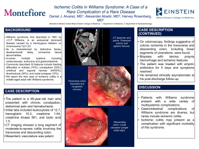
Case Description/Methods: The patient is a 48-year-old man who presented with chronic constipation, abdominal pain and hematochezia. Initial labs included leukocytosis of 12.7, hemoglobin 15.2, creatinine 1.64, CK 561, and lactic acid 1.5. CT imaging showed a long segment of moderate-to-severe colitis involving the transverse and descending colon (figure 1-A and 1-B). Mesenteric vasculature was normal. On colonoscopy, findings suggestive of colonic ischemia in the transverse and descending colon, including linear segments of ulcerations, were found (figure 1-C and 1-D). Biopsies with lamina propria hemorrhage and ischemic features. He was treated with antibiotics for 5 days and symptoms resolved. He remained clinically asymptomatic at his post-discharge follow-up.
Discussion: Patients with Williams syndrome present with a wide variety of multisystemic complications. Gastrointestinal complications of Williams syndrome are diverse, but rarely include ischemic colitis. Ischemic colitis may present as a complication with significant morbidity of this syndrome.

Disclosures:
Daniel J. Alvarez, MD, Alexander Abadir, MD, Harvey Rosenberg, MD. P0282 - Ischemic Colitis in Williams Syndrome: A Case of a Rare Complication of a Rare Disease, ACG 2023 Annual Scientific Meeting Abstracts. Vancouver, BC, Canada: American College of Gastroenterology.
Montefiore Medical Center/Albert Einstein College of Medicine, Bronx, NY
Introduction: Williams syndrome, first described in 1961 by J.C.P Williams, is an autosomal dominant disease caused by a hemizygous deletion on chromosome 7q11.23. This syndrome, with an estimated prevalence of 1:7,500 to 1:20,000, is characterized by distinctive facies, developmental delay, connective tissue abnormalities and involves multiple systems including cardiovascular, endocrine and gastrointestinal. Commonly described gastrointestinal features include feeding difficulties in children (70%), constipation (50%), umbilical and inguinal hernias (40/50%), diverticulosis (30%), and rectal prolapse (15%). We report the rare case of ischemic colitis in a middle-aged adult with Williams syndrome.
Case Description/Methods: The patient is a 48-year-old man who presented with chronic constipation, abdominal pain and hematochezia. Initial labs included leukocytosis of 12.7, hemoglobin 15.2, creatinine 1.64, CK 561, and lactic acid 1.5. CT imaging showed a long segment of moderate-to-severe colitis involving the transverse and descending colon (figure 1-A and 1-B). Mesenteric vasculature was normal. On colonoscopy, findings suggestive of colonic ischemia in the transverse and descending colon, including linear segments of ulcerations, were found (figure 1-C and 1-D). Biopsies with lamina propria hemorrhage and ischemic features. He was treated with antibiotics for 5 days and symptoms resolved. He remained clinically asymptomatic at his post-discharge follow-up.
Discussion: Patients with Williams syndrome present with a wide variety of multisystemic complications. Gastrointestinal complications of Williams syndrome are diverse, but rarely include ischemic colitis. Ischemic colitis may present as a complication with significant morbidity of this syndrome.

Figure: A: CT abdomen and pelvis: Thickened colonic wall (splenic flexure).
B: CT abdomen and pelvis: Thickened colonic wall (transverse and descending colon).
C: Transverse colon: Ulcerated and congested mucosa.
D: Descending colon: Linear ulceration (single-stripe sign).
B: CT abdomen and pelvis: Thickened colonic wall (transverse and descending colon).
C: Transverse colon: Ulcerated and congested mucosa.
D: Descending colon: Linear ulceration (single-stripe sign).
Disclosures:
Daniel Alvarez indicated no relevant financial relationships.
Alexander Abadir indicated no relevant financial relationships.
Harvey Rosenberg indicated no relevant financial relationships.
Daniel J. Alvarez, MD, Alexander Abadir, MD, Harvey Rosenberg, MD. P0282 - Ischemic Colitis in Williams Syndrome: A Case of a Rare Complication of a Rare Disease, ACG 2023 Annual Scientific Meeting Abstracts. Vancouver, BC, Canada: American College of Gastroenterology.
