Sunday Poster Session
Category: Colon
P0283 - The Clue in the Toilet: A Surprising Discovery of Gynecological Cancer Through Unlikely Symptoms
Sunday, October 22, 2023
3:30 PM - 7:00 PM PT
Location: Exhibit Hall

Has Audio
- DR
Diego Román-Colón, MD
VA Caribbean Healthcare System
San Juan, PR
Presenting Author(s)
Diego Román-Colón, MD, Esteban Grovas-Cordovi, MD, Alexander Alfaro, MD, Carla Blanco, MD, Priscilla Magno, MD, MSc, Jaime Martinez-Souss, MD
VA Caribbean Healthcare System, San Juan, Puerto Rico
Introduction: Diarrheal disease constitutes an important cause of medical admissions and deaths worldwide. Although common, the etiology, clinical presentation and management of these conditions can be broad and sometimes challenging. We present a case of chronic non-bloody diarrhea in which a multidisciplinary approach allowed an effective diagnosis and proper management of the condition.
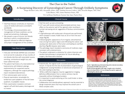
Case Description/Methods: Case of a 66-year-old female G0P0A0 that was consulted to our service for evaluation of two months of non-bloody diarrhea with associated nausea, vomiting, unintentional weight loss and lower abdominal pain. The patient had history of a total abdominal hysterectomy with bilateral salpingo-oophorectomy for suspected ovarian malignancy, although the pathology revealed endometriosis. Prior colonoscopies without polyps. Diarrhea workup was unrevealing. Physical examination remarkable for tenderness upon palpation of the left lower quadrant, no palpable masses. Pelvic examination revealed a remnant vaginal cuff with fetid watery discharge and no masses. Further imaging studies were recommended. A transvaginal sonogram demonstrated a pelvic soft tissue mass arising from the vaginal cuff, inseparable from the adjacent large bowel with a fistulous connection. A sigmoidoscopy with endoscopic ultrasound was performed which showed a raised, friable mass at 10 cm from the anal verge with endosonographic appearance of an irregular, extraintestinal mass arising from gynecological region and infiltrating the colonic wall causing near obstruction of the lumen and leading to overflow diarrhea, biopsies were taken. The pathology report revealed a carcinoma of mullerian origin consistent with endometrioma. The patient underwent a colostomy for colonic diversion and started chemotherapy in preparation for future debulking surgery, diarrheas were resolved.
Discussion: Anatomically, the vaginal vault is located posterior to the rectum. It may be the first site of presentation of gynecological malignancies causing fistulation or extramural compression of the colon leading to diarrhea, as in our case. While a definitive diagnosis may be suggested on imaging, definitive differentiation from a colonic primary may be impossible without histological analysis. It is important to avoid anchoring as this could hinder or delay the correct diagnosis. Therefore, collaboration and interdisciplinary approach may be essential to design treatment plans that may improve the overall care in these complex cases.

Disclosures:
Diego Román-Colón, MD, Esteban Grovas-Cordovi, MD, Alexander Alfaro, MD, Carla Blanco, MD, Priscilla Magno, MD, MSc, Jaime Martinez-Souss, MD. P0283 - The Clue in the Toilet: A Surprising Discovery of Gynecological Cancer Through Unlikely Symptoms, ACG 2023 Annual Scientific Meeting Abstracts. Vancouver, BC, Canada: American College of Gastroenterology.
VA Caribbean Healthcare System, San Juan, Puerto Rico
Introduction: Diarrheal disease constitutes an important cause of medical admissions and deaths worldwide. Although common, the etiology, clinical presentation and management of these conditions can be broad and sometimes challenging. We present a case of chronic non-bloody diarrhea in which a multidisciplinary approach allowed an effective diagnosis and proper management of the condition.
Case Description/Methods: Case of a 66-year-old female G0P0A0 that was consulted to our service for evaluation of two months of non-bloody diarrhea with associated nausea, vomiting, unintentional weight loss and lower abdominal pain. The patient had history of a total abdominal hysterectomy with bilateral salpingo-oophorectomy for suspected ovarian malignancy, although the pathology revealed endometriosis. Prior colonoscopies without polyps. Diarrhea workup was unrevealing. Physical examination remarkable for tenderness upon palpation of the left lower quadrant, no palpable masses. Pelvic examination revealed a remnant vaginal cuff with fetid watery discharge and no masses. Further imaging studies were recommended. A transvaginal sonogram demonstrated a pelvic soft tissue mass arising from the vaginal cuff, inseparable from the adjacent large bowel with a fistulous connection. A sigmoidoscopy with endoscopic ultrasound was performed which showed a raised, friable mass at 10 cm from the anal verge with endosonographic appearance of an irregular, extraintestinal mass arising from gynecological region and infiltrating the colonic wall causing near obstruction of the lumen and leading to overflow diarrhea, biopsies were taken. The pathology report revealed a carcinoma of mullerian origin consistent with endometrioma. The patient underwent a colostomy for colonic diversion and started chemotherapy in preparation for future debulking surgery, diarrheas were resolved.
Discussion: Anatomically, the vaginal vault is located posterior to the rectum. It may be the first site of presentation of gynecological malignancies causing fistulation or extramural compression of the colon leading to diarrhea, as in our case. While a definitive diagnosis may be suggested on imaging, definitive differentiation from a colonic primary may be impossible without histological analysis. It is important to avoid anchoring as this could hinder or delay the correct diagnosis. Therefore, collaboration and interdisciplinary approach may be essential to design treatment plans that may improve the overall care in these complex cases.

Figure: CT with contrast (Images 1 & 2) and endoscopic view (Image 3) of endometrioid mass causing obstruction of colonic lumen.
Disclosures:
Diego Román-Colón indicated no relevant financial relationships.
Esteban Grovas-Cordovi indicated no relevant financial relationships.
Alexander Alfaro indicated no relevant financial relationships.
Carla Blanco indicated no relevant financial relationships.
Priscilla Magno indicated no relevant financial relationships.
Jaime Martinez-Souss indicated no relevant financial relationships.
Diego Román-Colón, MD, Esteban Grovas-Cordovi, MD, Alexander Alfaro, MD, Carla Blanco, MD, Priscilla Magno, MD, MSc, Jaime Martinez-Souss, MD. P0283 - The Clue in the Toilet: A Surprising Discovery of Gynecological Cancer Through Unlikely Symptoms, ACG 2023 Annual Scientific Meeting Abstracts. Vancouver, BC, Canada: American College of Gastroenterology.
