Sunday Poster Session
Category: Biliary/Pancreas
P0057 - IgG4-Related Sclerosing Cholangitis Mimicking a Klatskin Tumor: Take Caution Before Resection
Sunday, October 22, 2023
3:30 PM - 7:00 PM PT
Location: Exhibit Hall

Has Audio

Laura Suzanne K. Suarez, MD
University of Florida-Jacksonville
Jacksonville, FL
Presenting Author(s)
Laura Suzanne K.. Suarez, MD1, Amit Sah, MD2, Ron Schey, MD1
1University of Florida-Jacksonville, Jacksonville, FL; 2University of Miami, JFK Medical Center, Atlantis, FL
Introduction: A Klatskin tumor or Perihilar Cholangiocarcinoma is a rare malignancy that occurs at the extrahepatic bile duct bifurcation. Tissue diagnosis is challenging due to the risk of tumor seeding. Due to its high mortality, if clinical and radiographic findings are sufficiently supportive of cholangiocarcinoma, biopsy prior to surgery is often not pursued. This approach remains to have caveats as we present a patient who underwent radical surgical resection of a presumed Klatskin tumor and was eventually diagnosed with IgG4-related sclerosing cholangitis.
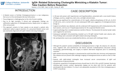
Case Description/Methods: A 59-year-old woman with hypertension and hypothyroidism, presented with a one-month history of fatigue, pruritus, weight loss, dark urine, and light-colored stools. Work-up was significant for a total bilirubin of 19.3 mg/dL, alkaline phosphatase of 191 u/L, Ca 19-9 of 196 U/mL, and a normal CEA. US and subesequent MRI were suggestive of cholangiocarcinoma (Image 1). Surgical consultation was obtained, and she underwent left hepatectomy with cholecystectomy, bile duct excision, and roux-en-Y hepaticojejunostomy for the suspected Klatskin tumor. Surgical pathology then revealed extensive storiform fibrosis. The dissected 18 reactive lymph nodes were negative for carcinoma. Subsequent IgG/IgG4 immunostaining revealed up to 30 IgG4 stained focally seen plasma cells. These findings were consistent with IgG4-related sclerosing cholangitis. Her postoperative course was complicated by biloma, and sepsis with multiorgan failure.
Discussion: Although this patient’s pretest probability of cholangiocarcinoma is high, the absence of a discreet mass, negative CEA, and autoimmune risk factors warrants multidisciplinary discussion among hematology, gastroenterology, and surgery to explore alternative diagnoses prior to considering resection. IgG4-related sclerosing cholangitis is characterized by multi-focal bile duct strictures and progressive liver disease and can present with radiologic imaging of cholangiocarcinoma as well as a liver or hilar mass. Patients with IgG4-related cholangitis have increased serum concentrations of IgG4 and responsiveness to glucocorticoids. All patients suspected of cholangiocarcinoma should have Ca-19-9, CEA, and AFP checked. Routinely checking IgG4 remains unclear. Appropriate staging can be done with EUS for patients with extrahepatic bile duct dilation. If a mass is not adequately visualized on EUS, then cholangioscopy or CT/PET is beneficial.

Disclosures:
Laura Suzanne K.. Suarez, MD1, Amit Sah, MD2, Ron Schey, MD1. P0057 - IgG4-Related Sclerosing Cholangitis Mimicking a Klatskin Tumor: Take Caution Before Resection, ACG 2023 Annual Scientific Meeting Abstracts. Vancouver, BC, Canada: American College of Gastroenterology.
1University of Florida-Jacksonville, Jacksonville, FL; 2University of Miami, JFK Medical Center, Atlantis, FL
Introduction: A Klatskin tumor or Perihilar Cholangiocarcinoma is a rare malignancy that occurs at the extrahepatic bile duct bifurcation. Tissue diagnosis is challenging due to the risk of tumor seeding. Due to its high mortality, if clinical and radiographic findings are sufficiently supportive of cholangiocarcinoma, biopsy prior to surgery is often not pursued. This approach remains to have caveats as we present a patient who underwent radical surgical resection of a presumed Klatskin tumor and was eventually diagnosed with IgG4-related sclerosing cholangitis.
Case Description/Methods: A 59-year-old woman with hypertension and hypothyroidism, presented with a one-month history of fatigue, pruritus, weight loss, dark urine, and light-colored stools. Work-up was significant for a total bilirubin of 19.3 mg/dL, alkaline phosphatase of 191 u/L, Ca 19-9 of 196 U/mL, and a normal CEA. US and subesequent MRI were suggestive of cholangiocarcinoma (Image 1). Surgical consultation was obtained, and she underwent left hepatectomy with cholecystectomy, bile duct excision, and roux-en-Y hepaticojejunostomy for the suspected Klatskin tumor. Surgical pathology then revealed extensive storiform fibrosis. The dissected 18 reactive lymph nodes were negative for carcinoma. Subsequent IgG/IgG4 immunostaining revealed up to 30 IgG4 stained focally seen plasma cells. These findings were consistent with IgG4-related sclerosing cholangitis. Her postoperative course was complicated by biloma, and sepsis with multiorgan failure.
Discussion: Although this patient’s pretest probability of cholangiocarcinoma is high, the absence of a discreet mass, negative CEA, and autoimmune risk factors warrants multidisciplinary discussion among hematology, gastroenterology, and surgery to explore alternative diagnoses prior to considering resection. IgG4-related sclerosing cholangitis is characterized by multi-focal bile duct strictures and progressive liver disease and can present with radiologic imaging of cholangiocarcinoma as well as a liver or hilar mass. Patients with IgG4-related cholangitis have increased serum concentrations of IgG4 and responsiveness to glucocorticoids. All patients suspected of cholangiocarcinoma should have Ca-19-9, CEA, and AFP checked. Routinely checking IgG4 remains unclear. Appropriate staging can be done with EUS for patients with extrahepatic bile duct dilation. If a mass is not adequately visualized on EUS, then cholangioscopy or CT/PET is beneficial.

Figure: Magnetic resonance imaging revealed significant intrahepatic and extrahepatic biliary ductal dilation and no discreet mass.
Disclosures:
Laura Suzanne Suarez indicated no relevant financial relationships.
Amit Sah indicated no relevant financial relationships.
Ron Schey indicated no relevant financial relationships.
Laura Suzanne K.. Suarez, MD1, Amit Sah, MD2, Ron Schey, MD1. P0057 - IgG4-Related Sclerosing Cholangitis Mimicking a Klatskin Tumor: Take Caution Before Resection, ACG 2023 Annual Scientific Meeting Abstracts. Vancouver, BC, Canada: American College of Gastroenterology.
