Sunday Poster Session
Category: Biliary/Pancreas
P0058 - Paraneoplastic Myositis as a Presenting Symptom of Pancreatic Adenocarcinoma
Sunday, October 22, 2023
3:30 PM - 7:00 PM PT
Location: Exhibit Hall

Has Audio

Gianna Stoleru, MD
University of Virginia
Charlottesville, VA
Presenting Author(s)
Gianna Stoleru, MD, Mark Radlinski, MD, Akriti Gupta, MD, Daniel Strand, MD
University of Virginia, Charlottesville, VA
Introduction: A 68-year-old man was admitted to the hospital for bilateral thigh weakness and found to have a pancreatic adenocarcinoma. This vignette highlights the heterogeneity of paraneoplastic syndromes and importance of multidisciplinary consultation.
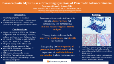
Case Description/Methods: A 68-year-old male with type 2 diabetes mellitus presented to the hospital with profound bilateral thigh weakness and malaise. He reported unintentional weight loss, dark urine, and scleral icterus over the 3 weeks preceding hospital presentation. Laboratory analysis revealed alkaline phosphatase 1388 U/L, total bilirubin 14.9 mg/dL (direct bilirubin 11 mg/dL), AST 916 U/L, ALT 338 U/L, INR 1.6, CK 31000 U/L, CA 19-9 199 U/mL, LDH 1669U/L. Abdominal ultrasound demonstrated intra- and extrahepatic biliary ductal dilation and a markedly enlarged pancreatic duct (PD). Subsequent (motion-limited) MRI/MRCP with and without contrast revealed an ill-defined intermediate T2 signal and mild restricted diffusion near the PD head suggestive of underlying malignancy. MRI of the thighs showed patchy intramuscular edema, suggestive of nonspecific myositis. Diagnostic EGD and EUS were performed which demonstrated diffuse features of advanced chronic calcific pancreatitis, including a ventral duct stone complex, biliary stricture and tissue expansion in the pancreatic head concerning for neoplasia. EUS directed FNA of the pancreatic head was performed, confirming adenocarcinoma. Concurrent ERCP demonstrated a high-grade mid-bile duct stricture with marked upstream dilation. Endoscopic biliary and pancreatic sphincterotomies were successful, followed by placement of a prophylactic ventral PD stent and fully covered metal biliary stent. Rheumatology was consulted for further management of his myositis. Muscle biopsy showed a nonspecific severe necrotizing myopathic process. Corticosteroids were initiated with subsequent downtrend in CK. The Oncology service recommended systemic chemotherapy given the lack of a discrete lesion amenable to resection.
Discussion: Very few case reports describe myositis as a presenting symptom of pancreatic adenocarcinoma. Common presenting symptoms in these cases included muscle weakness, generalized fatigue, and weight loss. Pathogenesis is thought to include a tumor driven, but subsequently self-perpetuating, immune response against muscle antigens. Corticosteroids and treatment of the underlying malignancy are the mainstay of therapy for paraneoplastic myositis.

Disclosures:
Gianna Stoleru, MD, Mark Radlinski, MD, Akriti Gupta, MD, Daniel Strand, MD. P0058 - Paraneoplastic Myositis as a Presenting Symptom of Pancreatic Adenocarcinoma, ACG 2023 Annual Scientific Meeting Abstracts. Vancouver, BC, Canada: American College of Gastroenterology.
University of Virginia, Charlottesville, VA
Introduction: A 68-year-old man was admitted to the hospital for bilateral thigh weakness and found to have a pancreatic adenocarcinoma. This vignette highlights the heterogeneity of paraneoplastic syndromes and importance of multidisciplinary consultation.
Case Description/Methods: A 68-year-old male with type 2 diabetes mellitus presented to the hospital with profound bilateral thigh weakness and malaise. He reported unintentional weight loss, dark urine, and scleral icterus over the 3 weeks preceding hospital presentation. Laboratory analysis revealed alkaline phosphatase 1388 U/L, total bilirubin 14.9 mg/dL (direct bilirubin 11 mg/dL), AST 916 U/L, ALT 338 U/L, INR 1.6, CK 31000 U/L, CA 19-9 199 U/mL, LDH 1669U/L. Abdominal ultrasound demonstrated intra- and extrahepatic biliary ductal dilation and a markedly enlarged pancreatic duct (PD). Subsequent (motion-limited) MRI/MRCP with and without contrast revealed an ill-defined intermediate T2 signal and mild restricted diffusion near the PD head suggestive of underlying malignancy. MRI of the thighs showed patchy intramuscular edema, suggestive of nonspecific myositis. Diagnostic EGD and EUS were performed which demonstrated diffuse features of advanced chronic calcific pancreatitis, including a ventral duct stone complex, biliary stricture and tissue expansion in the pancreatic head concerning for neoplasia. EUS directed FNA of the pancreatic head was performed, confirming adenocarcinoma. Concurrent ERCP demonstrated a high-grade mid-bile duct stricture with marked upstream dilation. Endoscopic biliary and pancreatic sphincterotomies were successful, followed by placement of a prophylactic ventral PD stent and fully covered metal biliary stent. Rheumatology was consulted for further management of his myositis. Muscle biopsy showed a nonspecific severe necrotizing myopathic process. Corticosteroids were initiated with subsequent downtrend in CK. The Oncology service recommended systemic chemotherapy given the lack of a discrete lesion amenable to resection.
Discussion: Very few case reports describe myositis as a presenting symptom of pancreatic adenocarcinoma. Common presenting symptoms in these cases included muscle weakness, generalized fatigue, and weight loss. Pathogenesis is thought to include a tumor driven, but subsequently self-perpetuating, immune response against muscle antigens. Corticosteroids and treatment of the underlying malignancy are the mainstay of therapy for paraneoplastic myositis.

Figure: A. Cytology of pancreatic mass (Papaniculaou stain, x400). The fine needle aspirate of the tumor showed clusters of malignant cells with marked nuclear size variation, irregular nuclear membranes, and fine cytoplasm.
B. Muscle biopsy (H&E, x200). The biopsy from the muscle demonstrates necrotic fibers (black arrow) with no significant inflammatory infiltrate.
C. Muscle biopsy (H&E, x400). Multinucleated macrophage (orange arrow) phagocytosing the necrotic muscle fiber (black arrow).
B. Muscle biopsy (H&E, x200). The biopsy from the muscle demonstrates necrotic fibers (black arrow) with no significant inflammatory infiltrate.
C. Muscle biopsy (H&E, x400). Multinucleated macrophage (orange arrow) phagocytosing the necrotic muscle fiber (black arrow).
Disclosures:
Gianna Stoleru indicated no relevant financial relationships.
Mark Radlinski indicated no relevant financial relationships.
Akriti Gupta indicated no relevant financial relationships.
Daniel Strand indicated no relevant financial relationships.
Gianna Stoleru, MD, Mark Radlinski, MD, Akriti Gupta, MD, Daniel Strand, MD. P0058 - Paraneoplastic Myositis as a Presenting Symptom of Pancreatic Adenocarcinoma, ACG 2023 Annual Scientific Meeting Abstracts. Vancouver, BC, Canada: American College of Gastroenterology.
