Sunday Poster Session
Category: Biliary/Pancreas
P0065 - Gallstone Ileus
Sunday, October 22, 2023
3:30 PM - 7:00 PM PT
Location: Exhibit Hall

Has Audio

Kathryn Evey, DO
Warren Alpert Medical School of Brown University
Providence, RI
Presenting Author(s)
Kathryn Evey, DO1, Andrew Krane, MD1, Sean Fine, MD, MS2
1Warren Alpert Medical School of Brown University, Providence, RI; 2Warren Alpert Medical School of Brown University, Providence, RI
Introduction: Of patients who present to the emergency room for abdominal pain, 2% have a small bowel obstruction (SBO), and only 4% of all SBOs are caused by gallstone ileus. This occurs when a gallstone travels through a fistula into the small intestine, causing obstruction. Gallstone ileus is associated with a high mortality rate (12-27%), due to delayed diagnosis and intervention. The gold standard for diagnosis is abdominopelvic CT imaging, and standard treatment is surgical intervention with stone removal. Here we present a case of gallstone ileus secondary to cholecystoduodenal fistula.
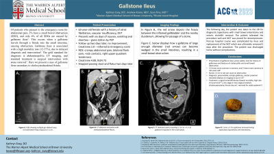
Case Description/Methods: A 63-year-old female with a past medical history of atrial fibrillation presented to the hospital after six days of nausea, vomiting and diarrhea. She reported close sick contacts with similar gastrointestinal complaints. Physical exam was notable for right upper quadrant tenderness to palpation. Laboratory analysis was notable for a creatinine of 4.89 and BUN 73. She was admitted for presumed viral enteritis and acute kidney injury. Two days later, she could no longer pass stool or flatus. Abdominal x-ray noted multiple gas-dilated loops of small intestine (Figure 1, A), concerning for SBO. Subsequent computed tomography scan revealed a cholecystoduodenal fistula (Figure 1, B) and a large gallstone impacted in the ileum (Figure 1, C). The patient underwent exploratory laparotomy and enterotomy, removing a 1.6cm x 3.7cm gallstone (Figure 1, D) from her jejunum. She tolerated the procedure well and was discharged home four days later without complications.
Discussion: Gallstone ileus (GI) is caused by cholecystitis that facilitates bilienteric adhesions, later forming a fistula for passage of a cholelith. In order to cause obstruction of the small bowel, stones frequently measure >2 cm in diameter. GI is seen more frequently in women (3.5:1), and the incidence is 4% in patients under 65 years old and 25% in patients over 65 years old with SBO. Diagnosis is crucial in identifying and expediting intervention, but its presentation varies widely. Cholecystoduodenal fistula is the most common type of fistula developed, as in our case, but fistula can form anywhere along the gastrointestinal tract. Enterolithotomy is the most common surgical procedure achieving clinical cure with minimal operative time and the lowest mortality, but it has higher rates of recurrence. Our case highlights the difficulties in initially diagnosing this entity and the importance of prompt surgical intervention.

Disclosures:
Kathryn Evey, DO1, Andrew Krane, MD1, Sean Fine, MD, MS2. P0065 - Gallstone Ileus, ACG 2023 Annual Scientific Meeting Abstracts. Vancouver, BC, Canada: American College of Gastroenterology.
1Warren Alpert Medical School of Brown University, Providence, RI; 2Warren Alpert Medical School of Brown University, Providence, RI
Introduction: Of patients who present to the emergency room for abdominal pain, 2% have a small bowel obstruction (SBO), and only 4% of all SBOs are caused by gallstone ileus. This occurs when a gallstone travels through a fistula into the small intestine, causing obstruction. Gallstone ileus is associated with a high mortality rate (12-27%), due to delayed diagnosis and intervention. The gold standard for diagnosis is abdominopelvic CT imaging, and standard treatment is surgical intervention with stone removal. Here we present a case of gallstone ileus secondary to cholecystoduodenal fistula.
Case Description/Methods: A 63-year-old female with a past medical history of atrial fibrillation presented to the hospital after six days of nausea, vomiting and diarrhea. She reported close sick contacts with similar gastrointestinal complaints. Physical exam was notable for right upper quadrant tenderness to palpation. Laboratory analysis was notable for a creatinine of 4.89 and BUN 73. She was admitted for presumed viral enteritis and acute kidney injury. Two days later, she could no longer pass stool or flatus. Abdominal x-ray noted multiple gas-dilated loops of small intestine (Figure 1, A), concerning for SBO. Subsequent computed tomography scan revealed a cholecystoduodenal fistula (Figure 1, B) and a large gallstone impacted in the ileum (Figure 1, C). The patient underwent exploratory laparotomy and enterotomy, removing a 1.6cm x 3.7cm gallstone (Figure 1, D) from her jejunum. She tolerated the procedure well and was discharged home four days later without complications.
Discussion: Gallstone ileus (GI) is caused by cholecystitis that facilitates bilienteric adhesions, later forming a fistula for passage of a cholelith. In order to cause obstruction of the small bowel, stones frequently measure >2 cm in diameter. GI is seen more frequently in women (3.5:1), and the incidence is 4% in patients under 65 years old and 25% in patients over 65 years old with SBO. Diagnosis is crucial in identifying and expediting intervention, but its presentation varies widely. Cholecystoduodenal fistula is the most common type of fistula developed, as in our case, but fistula can form anywhere along the gastrointestinal tract. Enterolithotomy is the most common surgical procedure achieving clinical cure with minimal operative time and the lowest mortality, but it has higher rates of recurrence. Our case highlights the difficulties in initially diagnosing this entity and the importance of prompt surgical intervention.

Figure: A. Upright Abdominal X-ray, demonstrating dilated loops of small bowel. B. CT Abdomen Pelvis, Coronal view, with cholecystoduodenal fistula (arrow). C. CT Abdomen Pelvis, Coronal view, gallstone impacted in distal jejunum. D. 1.6cm x 3.7cm gallstone after enterolithotomy.
Disclosures:
Kathryn Evey indicated no relevant financial relationships.
Andrew Krane indicated no relevant financial relationships.
Sean Fine: AbbVie – Advisory Committee/Board Member, Consultant. Bristol Myers Squibb – Advisory Committee/Board Member, Consultant.
Kathryn Evey, DO1, Andrew Krane, MD1, Sean Fine, MD, MS2. P0065 - Gallstone Ileus, ACG 2023 Annual Scientific Meeting Abstracts. Vancouver, BC, Canada: American College of Gastroenterology.
