Monday Poster Session
Category: Esophagus
P1818 - Artificial Intelligence and Capsule Panendoscopy: Development of a Convolutional Neural Network for the Automatic Detection of Pleomorphic Esophageal Lesions
Monday, October 23, 2023
10:30 AM - 4:15 PM PT
Location: Exhibit Hall

Has Audio
- TR
Tiago Ribeiro, MD
Centro Hospitalar São João
Porto, Porto, Portugal
Presenting Author(s)
Award: Presidential Poster Award
Tiago Ribeiro, MD1, Miguel Mascarenhas, MD2, João Afonso, MD1, Pedro Cardoso, MD2, Miguel Martins, MD2, Francisco Mendes, MD2, Patrícia Andrade, MD1, Hélder Cardoso, MD3, Miguel Saraiva, MD, PhD4, João Ferreira, PhD5, Guilherme Macedo, MD, PhD3
1Centro Hospitalar Universitário de São João, Porto, Porto, Portugal; 2Centro Hospitalar São João, Porto, Porto, Portugal; 3Centro Hospitalar de São João, Porto, Porto, Portugal; 4ManopH Gastroenterology Clinic, Porto, Porto, Portugal; 5University of Porto-FEUP, Porto, Porto, Portugal
Introduction: Capsule endoscopy (CE) has significantly improved the evaluation of the digestive tract, particularly the small intestine. In the recente years there has been a drive towards minimally-invasive capsule panendoscopy. Artificial intelligence (AI) algorithms are expected to mitigate the current limitations of CE and further stimulate the development of panendoscopy solutions. Nevertheless, although the esophagus is the first portion to be evaluated in CE exams, to date there is no evidence regarding the application of AI for the detection of esophageal lesions. The aim of this study was to develop the first AI model for the automatic detection of pleomorphic esophageal lesions.
Methods: A multicenter retrospective study was conducted with the aim of developing a Convolutional Neural Network (CNN) using 6977 esophageal images from 536 capsule endoscopy exams. The images were reviewed and validated by 3 CE specialists. Three different CE devices (Crohn's PillCam; PillCam SB3; OMOM HD capsule endoscopy system) were used to build the CNN. From the total number of included images, 3057 showed pleomorphic esophageal lesions and 3920 normal mucosa. The images were divided into training and validation groups, with 3 different evaluations being carried out to define the best model parameters. The performance of the model was ascertained by averaging the three evaluations, regarding its sensitivity, specificity, accuracy, positive predictive value (PPV), negative predictive value (NPV) and area under the precision-recall curve (AUPRC).
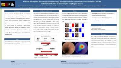
Results: Figure 1 depicts the heatmap showing the network’s perception of na esophageal lesion. The sensitivity, specificity, PPV and NPV were, respectively, 84.3%, 79.5%, 70.4%, 95.8%. The CNN achieved na accuracy of 84.2%, and an AUPRC of 0.942.
Discussion: This is the first study, worldwide, evaluating the performance of na AI model for the automatic detection of pleomorphic esophageal lesions using a minimally-invasive technique. The development of AI models specifically for the detection of esophageal lesions in CE is a necessary step to complete the implementation of AI-powered panendoscopic CE exams.

Disclosures:
Tiago Ribeiro, MD1, Miguel Mascarenhas, MD2, João Afonso, MD1, Pedro Cardoso, MD2, Miguel Martins, MD2, Francisco Mendes, MD2, Patrícia Andrade, MD1, Hélder Cardoso, MD3, Miguel Saraiva, MD, PhD4, João Ferreira, PhD5, Guilherme Macedo, MD, PhD3. P1818 - Artificial Intelligence and Capsule Panendoscopy: Development of a Convolutional Neural Network for the Automatic Detection of Pleomorphic Esophageal Lesions, ACG 2023 Annual Scientific Meeting Abstracts. Vancouver, BC, Canada: American College of Gastroenterology.
Tiago Ribeiro, MD1, Miguel Mascarenhas, MD2, João Afonso, MD1, Pedro Cardoso, MD2, Miguel Martins, MD2, Francisco Mendes, MD2, Patrícia Andrade, MD1, Hélder Cardoso, MD3, Miguel Saraiva, MD, PhD4, João Ferreira, PhD5, Guilherme Macedo, MD, PhD3
1Centro Hospitalar Universitário de São João, Porto, Porto, Portugal; 2Centro Hospitalar São João, Porto, Porto, Portugal; 3Centro Hospitalar de São João, Porto, Porto, Portugal; 4ManopH Gastroenterology Clinic, Porto, Porto, Portugal; 5University of Porto-FEUP, Porto, Porto, Portugal
Introduction: Capsule endoscopy (CE) has significantly improved the evaluation of the digestive tract, particularly the small intestine. In the recente years there has been a drive towards minimally-invasive capsule panendoscopy. Artificial intelligence (AI) algorithms are expected to mitigate the current limitations of CE and further stimulate the development of panendoscopy solutions. Nevertheless, although the esophagus is the first portion to be evaluated in CE exams, to date there is no evidence regarding the application of AI for the detection of esophageal lesions. The aim of this study was to develop the first AI model for the automatic detection of pleomorphic esophageal lesions.
Methods: A multicenter retrospective study was conducted with the aim of developing a Convolutional Neural Network (CNN) using 6977 esophageal images from 536 capsule endoscopy exams. The images were reviewed and validated by 3 CE specialists. Three different CE devices (Crohn's PillCam; PillCam SB3; OMOM HD capsule endoscopy system) were used to build the CNN. From the total number of included images, 3057 showed pleomorphic esophageal lesions and 3920 normal mucosa. The images were divided into training and validation groups, with 3 different evaluations being carried out to define the best model parameters. The performance of the model was ascertained by averaging the three evaluations, regarding its sensitivity, specificity, accuracy, positive predictive value (PPV), negative predictive value (NPV) and area under the precision-recall curve (AUPRC).
Results: Figure 1 depicts the heatmap showing the network’s perception of na esophageal lesion. The sensitivity, specificity, PPV and NPV were, respectively, 84.3%, 79.5%, 70.4%, 95.8%. The CNN achieved na accuracy of 84.2%, and an AUPRC of 0.942.
Discussion: This is the first study, worldwide, evaluating the performance of na AI model for the automatic detection of pleomorphic esophageal lesions using a minimally-invasive technique. The development of AI models specifically for the detection of esophageal lesions in CE is a necessary step to complete the implementation of AI-powered panendoscopic CE exams.

Figure: Figure 1. A - Capsule endoscopy image showing an esophageal protruding lesion. The heatmap (B) accurately identified the lesion.
Disclosures:
Tiago Ribeiro indicated no relevant financial relationships.
Miguel Mascarenhas indicated no relevant financial relationships.
João Afonso indicated no relevant financial relationships.
Pedro Cardoso indicated no relevant financial relationships.
Miguel Martins indicated no relevant financial relationships.
Francisco Mendes indicated no relevant financial relationships.
Patrícia Andrade indicated no relevant financial relationships.
Hélder Cardoso indicated no relevant financial relationships.
Miguel Saraiva indicated no relevant financial relationships.
João Ferreira indicated no relevant financial relationships.
Guilherme Macedo indicated no relevant financial relationships.
Tiago Ribeiro, MD1, Miguel Mascarenhas, MD2, João Afonso, MD1, Pedro Cardoso, MD2, Miguel Martins, MD2, Francisco Mendes, MD2, Patrícia Andrade, MD1, Hélder Cardoso, MD3, Miguel Saraiva, MD, PhD4, João Ferreira, PhD5, Guilherme Macedo, MD, PhD3. P1818 - Artificial Intelligence and Capsule Panendoscopy: Development of a Convolutional Neural Network for the Automatic Detection of Pleomorphic Esophageal Lesions, ACG 2023 Annual Scientific Meeting Abstracts. Vancouver, BC, Canada: American College of Gastroenterology.

