Monday Poster Session
Category: Esophagus
P1861 - Raise Your Anchor: The Importance of Ruling Out Malignancy in Cases of Suspected Achalasia
Monday, October 23, 2023
10:30 AM - 4:15 PM PT
Location: Exhibit Hall

Has Audio

Randy Leibowitz, DO
Mount Sinai Morningside and Mount Sinai West
New York, NY
Presenting Author(s)
Randy Leibowitz, DO1, Anudeep Neelam, MD2, Daniela Jodorkovsky, MD3
1Mount Sinai Morningside and Mount Sinai West, New York, NY; 2Mount Sinai Morningside West, New York, NY; 3Mount Sinai, New York, NY
Introduction: Pseudoachalasia can be difficult to differentiate from primary idiopathic achalasia. It is estimated 2-4% of patients who meet manometric criteria of achalasia have secondary disease from malignancy. On manometry, both cases can present with aperistalsis in the distal two-thirds of the esophagus and incomplete lower esophageal sphincter (LES) relaxation.
Case Description/Methods: A 76-year-old man with hypertension, dyslipidemia and type II diabetes presented with ongoing dysphagia, weight loss, and failure to thrive. He was admitted for further management. Dysphagia started four months prior to presentation and was only to solids. His dysphagia progressed rapidly to include liquids within a month of symptom onset. He endorsed 65 lb weight loss since the onset of symptoms.
He had previously presented to an outside hospital with recurrent dysphagia. He had undergone an EGD which was notable for a severe stenosis at the gastroesophageal junction (GEJ) which was dilated, and biopsies taken were negative for malignancy. The patient underwent high resolution manometry (HRM) which was consistent with type II achalasia. During a subsequent EGD, 100 units of botulinum injection were injected in the LES, however, the patient still did not have significant relief of symptoms.
He was scheduled to undergo Heller myotomy, however, he presented to our hospital with failure to thrive and dehydration associated with acute kidney injury. A plan was made to nutritionally optimize him with a nasoduodenal tube. However, during this EGD, the LES could not be traversed with a standard gastroscope and required an XP gastroscope. The cardia was nodular in appearance. He then underwent EUS showing a hypoechoic mass at the GEJ, lymphadenopathy, and pathology revealed a well-differentiated adenocarcinoma.
Discussion: Pseudoachalasia due to adenocarcinoma of the cardia could be due to mechanical obstruction of the GEJ or microscopic infiltration of the myenteric plexus or the vagus nerve. Presenting features associated with secondary achalasia due to malignancy include short duration of symptoms (i.e., less than six months), age greater than 60, excessive weight loss in relation to the duration of symptoms, and difficult passage of the endoscope through the GEJ.
In this case, anchor bias may have contributed to delay in detection and treatment of a gastric malignancy. Even if manometry shows classic features of achalasia, it is important to rule out pseudoachalasia in patients presenting with a rapidly progressive course.

Disclosures:
Randy Leibowitz, DO1, Anudeep Neelam, MD2, Daniela Jodorkovsky, MD3. P1861 - Raise Your Anchor: The Importance of Ruling Out Malignancy in Cases of Suspected Achalasia, ACG 2023 Annual Scientific Meeting Abstracts. Vancouver, BC, Canada: American College of Gastroenterology.
1Mount Sinai Morningside and Mount Sinai West, New York, NY; 2Mount Sinai Morningside West, New York, NY; 3Mount Sinai, New York, NY
Introduction: Pseudoachalasia can be difficult to differentiate from primary idiopathic achalasia. It is estimated 2-4% of patients who meet manometric criteria of achalasia have secondary disease from malignancy. On manometry, both cases can present with aperistalsis in the distal two-thirds of the esophagus and incomplete lower esophageal sphincter (LES) relaxation.
Case Description/Methods: A 76-year-old man with hypertension, dyslipidemia and type II diabetes presented with ongoing dysphagia, weight loss, and failure to thrive. He was admitted for further management. Dysphagia started four months prior to presentation and was only to solids. His dysphagia progressed rapidly to include liquids within a month of symptom onset. He endorsed 65 lb weight loss since the onset of symptoms.
He had previously presented to an outside hospital with recurrent dysphagia. He had undergone an EGD which was notable for a severe stenosis at the gastroesophageal junction (GEJ) which was dilated, and biopsies taken were negative for malignancy. The patient underwent high resolution manometry (HRM) which was consistent with type II achalasia. During a subsequent EGD, 100 units of botulinum injection were injected in the LES, however, the patient still did not have significant relief of symptoms.
He was scheduled to undergo Heller myotomy, however, he presented to our hospital with failure to thrive and dehydration associated with acute kidney injury. A plan was made to nutritionally optimize him with a nasoduodenal tube. However, during this EGD, the LES could not be traversed with a standard gastroscope and required an XP gastroscope. The cardia was nodular in appearance. He then underwent EUS showing a hypoechoic mass at the GEJ, lymphadenopathy, and pathology revealed a well-differentiated adenocarcinoma.
Discussion: Pseudoachalasia due to adenocarcinoma of the cardia could be due to mechanical obstruction of the GEJ or microscopic infiltration of the myenteric plexus or the vagus nerve. Presenting features associated with secondary achalasia due to malignancy include short duration of symptoms (i.e., less than six months), age greater than 60, excessive weight loss in relation to the duration of symptoms, and difficult passage of the endoscope through the GEJ.
In this case, anchor bias may have contributed to delay in detection and treatment of a gastric malignancy. Even if manometry shows classic features of achalasia, it is important to rule out pseudoachalasia in patients presenting with a rapidly progressive course.

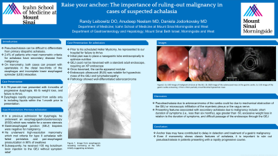
Figure: Image from esophagram revealing narrowing at the GEJ initially suggesting achalasia.
Disclosures:
Randy Leibowitz indicated no relevant financial relationships.
Anudeep Neelam indicated no relevant financial relationships.
Daniela Jodorkovsky: ATMO Biosciences – Consultant.
Randy Leibowitz, DO1, Anudeep Neelam, MD2, Daniela Jodorkovsky, MD3. P1861 - Raise Your Anchor: The Importance of Ruling Out Malignancy in Cases of Suspected Achalasia, ACG 2023 Annual Scientific Meeting Abstracts. Vancouver, BC, Canada: American College of Gastroenterology.
