Monday Poster Session
Category: Esophagus
P1881 - Beyond the Bone Marrow: Uncommon Manifestation of Multiple Myeloma - A Case Report of Extramedullary Esophageal Involvement
Monday, October 23, 2023
10:30 AM - 4:15 PM PT
Location: Exhibit Hall

Has Audio

Maya Mahmoud, MD
Saint Louis University
St. Louis, MO
Presenting Author(s)
Award: Presidential Poster Award
Maya Mahmoud, MD1, Mahmoud Y. Madi, MD2, Manar Shahwan, MD1, Eyad Gharaibeh, MD1, Ranju Kunwor, MD1, Wissam Kiwan, MD1, Antonio Cheesman, MD2
1Saint Louis University, St. Louis, MO; 2Saint Louis University Hospital, St. Louis, MO
Introduction: Extramedullary multiple myeloma is defined by the development of plasma cells outside the bone marrow. It is considered an aggressive subtype of multiple myeloma. Involvement of the gastrointestinal tract by plasma cells occurs in less than 5% of extramedullary myeloma.
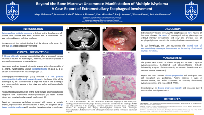
Case Description/Methods: We present the case of a 60-year-old male with a history of tobacco use who was admitted after a syncopal episode with facial trauma. Laboratory work-up showed microcytic anemia with a hemoglobin of 7.1 mg/dL, hypercalcemia and an incidental finding of 1.8 x 6.2 x 5.3 cm soft tissue lesion in the distal esophagus (Figure 1 A). Esophagogastroduodenoscopy (EGD) revealed a 5 cm, partially circumferential, friable, and ulcerated mass in the lower third of the esophagus (Fig. 1 B). Histopathological examination of the mass biopsy showed a hematolymphoid neoplasm with plasmacytic immunophenotype (Fig. 1 D). Bone marrow biopsy was negative for plasma cell involvement (Fig. 1 E). Positron emission tomography (PET) scan revealed a large avid mass in the esophagus, numerous lytic lesions in the calvarium, pelvis and appendicular skeleton (Figure 1 C). Based on esophagus pathology combined with serum M protein, anemia, hypercalcemia, and lytic lesions in bone the diagnosis of IgG Kappa multiple myeloma with standard risk cytogenetics is confirmed. The patient was started on chemotherapy and received 1 cycle of cyclophosphamide, bortezomib and dexamethasone (CyborD) followed by 3 cycles of daratumumab, bortezomib, lenalidomide and dexamethasone (DARA-VRd). Repeat PET scan revealed disease progression and autologous stem cell transplant was postponed. Patient received 1 cycle of dexamethasone, and 4-day continuous of cisplatin, doxorubicin, cyclophosphamide and etoposide (D-PACE). Unfortunately, his disease progressed rapidly, and he passed away 6 months after initial presentation.
Discussion: Our patient was diagnosed with extramedullary IgG kappa multiple myeloma with esophagus and bone involvement. Extramedullary lesions involving the esophagus are rare. Review of literature showed six cases of esophageal solitary plasmacytoma without marrow involvement, and only one previous case of esophageal plasmacytoma in the setting of a bone marrow disease. To our knowledge, our case represents the second case of extramedullary esophageal involvement in the setting of advanced multiple myeloma.

Disclosures:
Maya Mahmoud, MD1, Mahmoud Y. Madi, MD2, Manar Shahwan, MD1, Eyad Gharaibeh, MD1, Ranju Kunwor, MD1, Wissam Kiwan, MD1, Antonio Cheesman, MD2. P1881 - Beyond the Bone Marrow: Uncommon Manifestation of Multiple Myeloma - A Case Report of Extramedullary Esophageal Involvement, ACG 2023 Annual Scientific Meeting Abstracts. Vancouver, BC, Canada: American College of Gastroenterology.
Maya Mahmoud, MD1, Mahmoud Y. Madi, MD2, Manar Shahwan, MD1, Eyad Gharaibeh, MD1, Ranju Kunwor, MD1, Wissam Kiwan, MD1, Antonio Cheesman, MD2
1Saint Louis University, St. Louis, MO; 2Saint Louis University Hospital, St. Louis, MO
Introduction: Extramedullary multiple myeloma is defined by the development of plasma cells outside the bone marrow. It is considered an aggressive subtype of multiple myeloma. Involvement of the gastrointestinal tract by plasma cells occurs in less than 5% of extramedullary myeloma.
Case Description/Methods: We present the case of a 60-year-old male with a history of tobacco use who was admitted after a syncopal episode with facial trauma. Laboratory work-up showed microcytic anemia with a hemoglobin of 7.1 mg/dL, hypercalcemia and an incidental finding of 1.8 x 6.2 x 5.3 cm soft tissue lesion in the distal esophagus (Figure 1 A). Esophagogastroduodenoscopy (EGD) revealed a 5 cm, partially circumferential, friable, and ulcerated mass in the lower third of the esophagus (Fig. 1 B). Histopathological examination of the mass biopsy showed a hematolymphoid neoplasm with plasmacytic immunophenotype (Fig. 1 D). Bone marrow biopsy was negative for plasma cell involvement (Fig. 1 E). Positron emission tomography (PET) scan revealed a large avid mass in the esophagus, numerous lytic lesions in the calvarium, pelvis and appendicular skeleton (Figure 1 C). Based on esophagus pathology combined with serum M protein, anemia, hypercalcemia, and lytic lesions in bone the diagnosis of IgG Kappa multiple myeloma with standard risk cytogenetics is confirmed. The patient was started on chemotherapy and received 1 cycle of cyclophosphamide, bortezomib and dexamethasone (CyborD) followed by 3 cycles of daratumumab, bortezomib, lenalidomide and dexamethasone (DARA-VRd). Repeat PET scan revealed disease progression and autologous stem cell transplant was postponed. Patient received 1 cycle of dexamethasone, and 4-day continuous of cisplatin, doxorubicin, cyclophosphamide and etoposide (D-PACE). Unfortunately, his disease progressed rapidly, and he passed away 6 months after initial presentation.
Discussion: Our patient was diagnosed with extramedullary IgG kappa multiple myeloma with esophagus and bone involvement. Extramedullary lesions involving the esophagus are rare. Review of literature showed six cases of esophageal solitary plasmacytoma without marrow involvement, and only one previous case of esophageal plasmacytoma in the setting of a bone marrow disease. To our knowledge, our case represents the second case of extramedullary esophageal involvement in the setting of advanced multiple myeloma.

Figure: Figure 1
A: Computed tomography (CT) of the abdomen revealing a large, hyperattenuating, asymmetric soft tissue lesion within the distal thoracic esophagus measuring approximately 1.8 x 6.2 x 5.3 cm
B: Esophagogastroduodenoscopy (EGD) showing a friable, non-bleeding, partially circumferential, large, ulcerating mass in the lower third of the esophagus.
C: PET scan image pre-initiation of treatment: large avid mass in the esophagus, numerous lytic lesions calvarium, pelvis, appendicular skeleton.
D: Esophageal mass biopsy showing hematolymphoid neoplasm with plasmacytic immunophenotype.
E: Bone marrow with trilineage hematopoiesis and no evidence of plasma cell neoplasm.
Disclosures:
Maya Mahmoud indicated no relevant financial relationships.
Mahmoud Madi indicated no relevant financial relationships.
Manar Shahwan indicated no relevant financial relationships.
Eyad Gharaibeh indicated no relevant financial relationships.
Ranju Kunwor indicated no relevant financial relationships.
Wissam Kiwan indicated no relevant financial relationships.
Antonio Cheesman indicated no relevant financial relationships.
Maya Mahmoud, MD1, Mahmoud Y. Madi, MD2, Manar Shahwan, MD1, Eyad Gharaibeh, MD1, Ranju Kunwor, MD1, Wissam Kiwan, MD1, Antonio Cheesman, MD2. P1881 - Beyond the Bone Marrow: Uncommon Manifestation of Multiple Myeloma - A Case Report of Extramedullary Esophageal Involvement, ACG 2023 Annual Scientific Meeting Abstracts. Vancouver, BC, Canada: American College of Gastroenterology.

