Monday Poster Session
Category: Esophagus
P1883 - Sloughing Esophagitis: An Unexpected Incidental Finding
Monday, October 23, 2023
10:30 AM - 4:15 PM PT
Location: Exhibit Hall

Has Audio

Ayham Khrais, DO
Rutgers New Jersey Medical School
Newark, New Jersey
Presenting Author(s)
Ayham Khrais, DO, Hamza Khan, BS, Param Patel, MD, Ahmed Al-Khazraji, MD
Rutgers New Jersey Medical School, Newark, NJ
Introduction: Esophagitis dissecans superficialis (EDS) describes sloughing or desquamation of esophageal mucosa. It is rare, and often associated with mucosal injury and autoimmunity. Specifically, it has been correlated with dermatologic conditions such as bullous pemphigoid and pemphigus vulgaris. Patients can present with hematemesis, vomiting, dysphagia, odynophagia or they can be asymptomatic. Treatment involves removal of the offending agent and a course of proton pump inhibitors. Here, we present a case of EDS in a patient with a known granulomatous inflammatory nasal lesion who presented to the hospital with acute on chronic iron deficiency anemia.
Case Description/Methods: A 49-year-old female with a history of an inflammatory granulomatous nasal lesion and IDA secondary to bleeding uterine fibroids for which she underwent a hysterectomy presented with one week of lightheadedness and profuse bleeding from her nasal lesion after she had picked at it with a needle. Upon her arrival, she was hemodynamically stable, but was noted to have a hemoglobin of 3.9 g/dL. She was given 3 units of packed red blood cells with subsequent improvement in her hemoglobin to 8 g/dL. She underwent EGD to evaluate for an etiology of refractory iron deficiency anemia despite the use of iron supplementation. EGD revealed a white circumferential esophageal plaque suspicious for esophageal dissecans superficialis (Figure 1), as well as a healed clean-based ulcer in the gastric antrum without signs of bleeding. Pathology demonstrated mild basal cell hyperplasia with mildly elongated lamina propria papillae without significant inflammatory infiltrate, glandular epithelium, viral inclusions, fungal organisms or dysplasia. She was given an 8-week course of PPI and discharged with outpatient follow up.
Discussion: Here, we present a case of EDS in a patient with a known inflammatory skin lesion who presented with worsening iron deficiency anemia that was deemed likely secondary to bleeding from her skin lesion. The EDS did not provoke symptoms in our patient. Her EDS may be associated with her skin lesion, which has been biopsied but the significance of which is unknown.

Disclosures:
Ayham Khrais, DO, Hamza Khan, BS, Param Patel, MD, Ahmed Al-Khazraji, MD. P1883 - Sloughing Esophagitis: An Unexpected Incidental Finding, ACG 2023 Annual Scientific Meeting Abstracts. Vancouver, BC, Canada: American College of Gastroenterology.
Rutgers New Jersey Medical School, Newark, NJ
Introduction: Esophagitis dissecans superficialis (EDS) describes sloughing or desquamation of esophageal mucosa. It is rare, and often associated with mucosal injury and autoimmunity. Specifically, it has been correlated with dermatologic conditions such as bullous pemphigoid and pemphigus vulgaris. Patients can present with hematemesis, vomiting, dysphagia, odynophagia or they can be asymptomatic. Treatment involves removal of the offending agent and a course of proton pump inhibitors. Here, we present a case of EDS in a patient with a known granulomatous inflammatory nasal lesion who presented to the hospital with acute on chronic iron deficiency anemia.
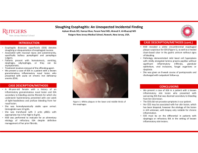
Case Description/Methods: A 49-year-old female with a history of an inflammatory granulomatous nasal lesion and IDA secondary to bleeding uterine fibroids for which she underwent a hysterectomy presented with one week of lightheadedness and profuse bleeding from her nasal lesion after she had picked at it with a needle. Upon her arrival, she was hemodynamically stable, but was noted to have a hemoglobin of 3.9 g/dL. She was given 3 units of packed red blood cells with subsequent improvement in her hemoglobin to 8 g/dL. She underwent EGD to evaluate for an etiology of refractory iron deficiency anemia despite the use of iron supplementation. EGD revealed a white circumferential esophageal plaque suspicious for esophageal dissecans superficialis (Figure 1), as well as a healed clean-based ulcer in the gastric antrum without signs of bleeding. Pathology demonstrated mild basal cell hyperplasia with mildly elongated lamina propria papillae without significant inflammatory infiltrate, glandular epithelium, viral inclusions, fungal organisms or dysplasia. She was given an 8-week course of PPI and discharged with outpatient follow up.
Discussion: Here, we present a case of EDS in a patient with a known inflammatory skin lesion who presented with worsening iron deficiency anemia that was deemed likely secondary to bleeding from her skin lesion. The EDS did not provoke symptoms in our patient. Her EDS may be associated with her skin lesion, which has been biopsied but the significance of which is unknown.

Figure: Figure 1: White Plaques in the Middle Third of the Esophagus.
Disclosures:
Ayham Khrais indicated no relevant financial relationships.
Hamza Khan indicated no relevant financial relationships.
Param Patel indicated no relevant financial relationships.
Ahmed Al-Khazraji indicated no relevant financial relationships.
Ayham Khrais, DO, Hamza Khan, BS, Param Patel, MD, Ahmed Al-Khazraji, MD. P1883 - Sloughing Esophagitis: An Unexpected Incidental Finding, ACG 2023 Annual Scientific Meeting Abstracts. Vancouver, BC, Canada: American College of Gastroenterology.
