Monday Poster Session
Category: Esophagus
P1920 - A Rare Case of Dysphagia Aortica Identified by Esophageal Manometry
Monday, October 23, 2023
10:30 AM - 4:15 PM PT
Location: Exhibit Hall

Has Audio

Megan B. Ghai, MD, MPH, MA
University of Arizona College of Medicine
Phoenix, AZ
Presenting Author(s)
Megan B. Ghai, MD, MPH, MA1, Natasha Narang, DO1, Rakesh Nanda, MD, FACG2
1University of Arizona College of Medicine, Phoenix, AZ; 2Phoenix VA Medical Center, Phoenix, AZ
Introduction: Dysphagia aortica is a rare condition in which a tortuous or dilated aorta compresses the esophagus causing dysphagia. There have only been 71 cases of dysphagia aortica documented in the literature. Presented is a rare case of dysphagia aortica caused by an aortic aneurysm that was identified on high-resolution esophageal manometry (HRM).
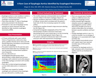
Case Description/Methods: A 70-year-old female with a past medical history of hypertension, obstructive sleep apnea, papillary thyroid cancer status post left thyroid lobectomy, and tobacco use presented to clinic with dysphagia. The patient stated that for the past two years food moved slowly down her esophagus, but she had started to noticed these symptoms more frequently. She was referred for an esophagogastroduodenoscopy which demonstrated a mildly dilated and tortuous esophagus with scattered round white plaques in the mid-esophagus consistent with Candida esophagitis. There was no observed gastroesophageal junction (GEJ) obstruction or resistance to scope. The patient was treated for Candida esophagitis yet continued to have dysphagia. She was referred for a motility study which was uninterpretable due to profound vascular artifact in the lower esophagus (Figure 1a). Vascular oscillations observed on HRM occurred at a rate of 108 per minute which correlated with the patient’s heart rate of 110, thus increasing suspicion for cardiac artifact. Timed barium swallow showed a large aneurysm abutting the GEJ (Figure 1b). Computed tomography angiography (CTa) of the chest demonstrated a fusiform aneurysm of the lower descending thoracic aorta measuring 4.3 cm but increasing to 4.7 cm at the diaphragmatic hiatus (Figure 1c). The patient underwent endovascular repair of the aortic aneurysm with mild improvement of dysphagia.
Discussion: This is an unusual case of cardiac artifact on HRM that led to the diagnosis of dysphagia aortica. Falsely elevated high integrated relaxation pressures suggested an esophagogastric junction outflow obstruction thought to be due to cardiac artifact given the correlation between vascular oscillation and heart rates. Though dysphagia aortica is rare, it is critical to consider it as an etiology of dysphagia as it is associated with a high mortality rate. Those treated conservatively versus with surgical repair have reported mortality rates of 55% and 21%, respectively. If suspicion for dysphagia aortica is high, evaluation with a timed barium swallow and CTa can facilitate this diagnosis.

Disclosures:
Megan B. Ghai, MD, MPH, MA1, Natasha Narang, DO1, Rakesh Nanda, MD, FACG2. P1920 - A Rare Case of Dysphagia Aortica Identified by Esophageal Manometry, ACG 2023 Annual Scientific Meeting Abstracts. Vancouver, BC, Canada: American College of Gastroenterology.
1University of Arizona College of Medicine, Phoenix, AZ; 2Phoenix VA Medical Center, Phoenix, AZ
Introduction: Dysphagia aortica is a rare condition in which a tortuous or dilated aorta compresses the esophagus causing dysphagia. There have only been 71 cases of dysphagia aortica documented in the literature. Presented is a rare case of dysphagia aortica caused by an aortic aneurysm that was identified on high-resolution esophageal manometry (HRM).
Case Description/Methods: A 70-year-old female with a past medical history of hypertension, obstructive sleep apnea, papillary thyroid cancer status post left thyroid lobectomy, and tobacco use presented to clinic with dysphagia. The patient stated that for the past two years food moved slowly down her esophagus, but she had started to noticed these symptoms more frequently. She was referred for an esophagogastroduodenoscopy which demonstrated a mildly dilated and tortuous esophagus with scattered round white plaques in the mid-esophagus consistent with Candida esophagitis. There was no observed gastroesophageal junction (GEJ) obstruction or resistance to scope. The patient was treated for Candida esophagitis yet continued to have dysphagia. She was referred for a motility study which was uninterpretable due to profound vascular artifact in the lower esophagus (Figure 1a). Vascular oscillations observed on HRM occurred at a rate of 108 per minute which correlated with the patient’s heart rate of 110, thus increasing suspicion for cardiac artifact. Timed barium swallow showed a large aneurysm abutting the GEJ (Figure 1b). Computed tomography angiography (CTa) of the chest demonstrated a fusiform aneurysm of the lower descending thoracic aorta measuring 4.3 cm but increasing to 4.7 cm at the diaphragmatic hiatus (Figure 1c). The patient underwent endovascular repair of the aortic aneurysm with mild improvement of dysphagia.
Discussion: This is an unusual case of cardiac artifact on HRM that led to the diagnosis of dysphagia aortica. Falsely elevated high integrated relaxation pressures suggested an esophagogastric junction outflow obstruction thought to be due to cardiac artifact given the correlation between vascular oscillation and heart rates. Though dysphagia aortica is rare, it is critical to consider it as an etiology of dysphagia as it is associated with a high mortality rate. Those treated conservatively versus with surgical repair have reported mortality rates of 55% and 21%, respectively. If suspicion for dysphagia aortica is high, evaluation with a timed barium swallow and CTa can facilitate this diagnosis.

Figure: Figure 1a. Esophageal manometry with elevated high integrated relaxation pressures on HRM suggestive of an esophagogastric junction outflow obstruction (see arrow).
Figure 1b. Timed barium swallow shows a large aneurysm abutting the gastroesophageal junction (see arrow).
Figure 1c. Computed tomography angiography with a large aortic aneurysm compressing the esophagus (see arrow).
Figure 1b. Timed barium swallow shows a large aneurysm abutting the gastroesophageal junction (see arrow).
Figure 1c. Computed tomography angiography with a large aortic aneurysm compressing the esophagus (see arrow).
Disclosures:
Megan Ghai indicated no relevant financial relationships.
Natasha Narang indicated no relevant financial relationships.
Rakesh Nanda indicated no relevant financial relationships.
Megan B. Ghai, MD, MPH, MA1, Natasha Narang, DO1, Rakesh Nanda, MD, FACG2. P1920 - A Rare Case of Dysphagia Aortica Identified by Esophageal Manometry, ACG 2023 Annual Scientific Meeting Abstracts. Vancouver, BC, Canada: American College of Gastroenterology.
