Monday Poster Session
Category: Esophagus
P1921 - Gastroesophageal Junction Adenocarcinoma Presenting as a Shoulder Mass
Monday, October 23, 2023
10:30 AM - 4:15 PM PT
Location: Exhibit Hall

Has Audio

Benjamin W. Warren, MD
Houston Methodist Hospital
Houston, Texas
Presenting Author(s)
Benjamin W. Warren, MD, Ali Raza, MD
Houston Methodist Hospital, Houston, TX
Introduction: Esophageal cancer is the eighth-most common cancer worldwide. Prognosis is poor especially in the presence of distant metastasis. The most common sites of metastasis are lymph nodes, liver, and lung. We describe a case of metastatic gastroesophageal junction (GEJ) adenocarcinoma presenting as a left shoulder mass.
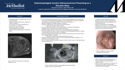
Case Description/Methods: A 76-year-old female presented to an orthopedic surgeon with seven months of severe, progressive left shoulder pain. Past medical history was significant for a remote cancer of the left axilla approximately 17 years prior for which she underwent left axillary lymph node resection and chemoradiation leading to chronic lymphedema of the left arm, however records were not available for review. A non-contrast MRI of the left shoulder demonstrated a large aggressive marrow replacing lesion centered in the glenoid with extension of the mass into the acromion, scapular spine, coracoid process, scapular body, rotator cuff musculature, glenohumeral joint, and humeral head. Differential diagnosis for these findings included metastatic disease versus myeloma. A computed tomography (CT) guided needle biopsy of the shoulder mass was performed, and pathology demonstrated metastatic adenocarcinoma. CT scan of the chest, abdomen and pelvis demonstrated the known shoulder lesion along with metastatic retroperitoneal adenopathy and indeterminate porta hepatis adenopathy. Gastroenterology was consulted to evaluate for a primary gastrointestinal (GI) malignancy. The patient denied any upper or lower GI symptoms including dysphagia, bleeding, or change in bowel habits. She had no prior colonoscopy. Esophagogastroduodenoscopy (EGD) revealed a large fungating, partially obstructing, partially circumferential mass in the lower third of the esophagus from which biopsies were obtained. Endoscopic ultrasound (EUS) of the mass demonstrated invasion into the muscularis propria (layer 4). The pancreas and liver appeared normal on EUS. Pathology from the esophageal mass demonstrated invasive poorly differentiated adenocarcinoma. She underwent palliative radiation therapy to the left shoulder mass but ultimately elected to pursue hospice care.
Discussion: Our patient’s presenting symptom was left shoulder pain due to a large underlying mass, ultimately determined to be metastasis from a GEJ adenocarcinoma. While bony metastasis from esophageal adenocarcinoma can occur, monoarticular metastasis to the glenohumeral joint has not been previously described.

Disclosures:
Benjamin W. Warren, MD, Ali Raza, MD. P1921 - Gastroesophageal Junction Adenocarcinoma Presenting as a Shoulder Mass, ACG 2023 Annual Scientific Meeting Abstracts. Vancouver, BC, Canada: American College of Gastroenterology.
Houston Methodist Hospital, Houston, TX
Introduction: Esophageal cancer is the eighth-most common cancer worldwide. Prognosis is poor especially in the presence of distant metastasis. The most common sites of metastasis are lymph nodes, liver, and lung. We describe a case of metastatic gastroesophageal junction (GEJ) adenocarcinoma presenting as a left shoulder mass.
Case Description/Methods: A 76-year-old female presented to an orthopedic surgeon with seven months of severe, progressive left shoulder pain. Past medical history was significant for a remote cancer of the left axilla approximately 17 years prior for which she underwent left axillary lymph node resection and chemoradiation leading to chronic lymphedema of the left arm, however records were not available for review. A non-contrast MRI of the left shoulder demonstrated a large aggressive marrow replacing lesion centered in the glenoid with extension of the mass into the acromion, scapular spine, coracoid process, scapular body, rotator cuff musculature, glenohumeral joint, and humeral head. Differential diagnosis for these findings included metastatic disease versus myeloma. A computed tomography (CT) guided needle biopsy of the shoulder mass was performed, and pathology demonstrated metastatic adenocarcinoma. CT scan of the chest, abdomen and pelvis demonstrated the known shoulder lesion along with metastatic retroperitoneal adenopathy and indeterminate porta hepatis adenopathy. Gastroenterology was consulted to evaluate for a primary gastrointestinal (GI) malignancy. The patient denied any upper or lower GI symptoms including dysphagia, bleeding, or change in bowel habits. She had no prior colonoscopy. Esophagogastroduodenoscopy (EGD) revealed a large fungating, partially obstructing, partially circumferential mass in the lower third of the esophagus from which biopsies were obtained. Endoscopic ultrasound (EUS) of the mass demonstrated invasion into the muscularis propria (layer 4). The pancreas and liver appeared normal on EUS. Pathology from the esophageal mass demonstrated invasive poorly differentiated adenocarcinoma. She underwent palliative radiation therapy to the left shoulder mass but ultimately elected to pursue hospice care.
Discussion: Our patient’s presenting symptom was left shoulder pain due to a large underlying mass, ultimately determined to be metastasis from a GEJ adenocarcinoma. While bony metastasis from esophageal adenocarcinoma can occur, monoarticular metastasis to the glenohumeral joint has not been previously described.

Figure: MRI demonstrating the left shoulder mass
Disclosures:
Benjamin Warren indicated no relevant financial relationships.
Ali Raza indicated no relevant financial relationships.
Benjamin W. Warren, MD, Ali Raza, MD. P1921 - Gastroesophageal Junction Adenocarcinoma Presenting as a Shoulder Mass, ACG 2023 Annual Scientific Meeting Abstracts. Vancouver, BC, Canada: American College of Gastroenterology.
