Monday Poster Session
Category: General Endoscopy
P1993 - Melanoma Leaves a Mark: Tumoral Melanosis of the Colon
Monday, October 23, 2023
10:30 AM - 4:15 PM PT
Location: Exhibit Hall

Has Audio
- AD
Anoushka Dua, MD
UCLA
Los Angeles, CA
Presenting Author(s)
Anoushka Dua, MD1, Tara Narasimhalu, MD2, Sepehr Hamidi, MD1, Rohit Dewan, DO1, Benjamin Nulsen, MD3
1UCLA, Los Angeles, CA; 2University of California, Los Angeles, Los Angeles, CA; 3David Geffen School of Medicine at UCLA, Los Angeles, CA
Introduction: Melanoma is one of the most common malignancies to metastasize to the GI tract. Any pigmented lesion visualized during colonoscopy in a patient with a history of melanoma should be biopsied to evaluate for intestinal metastases. However, tumoral melanosis is also on the differential. Tumoral melanosis is a rare, benign pigmented lesion representing a completely regressed melanoma, though strongly mimics malignant melanoma. It has been most commonly observed on the skin and lymph nodes — this represents the second documented case in the GI tract.
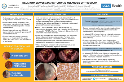
Case Description/Methods: A 63-year-old man with melanoma, metastatic at the time of diagnosis to the lungs, adrenal gland, small bowel, and colon and subsequently treated with 19 months of immunotherapy (nivolumab and ipilimumab followed by nivolumab monotherapy), presented for screening colonoscopy. PET-CT scan a few weeks prior to the colonoscopy demonstrated decreased colonic wall thickening and FDG uptake compared to PET-CT at the time of diagnosis, consistent with a favorable response to treatment. Colonoscopy revealed a suspicious-appearing 2cm pigmented lesion in the proximal ascending colon (A). Pathology of the biopsied colonic mucosa demonstrated a diffuse infiltrate of melanophages with abundant melanin and no evidence of melanoma (B), supported by immunohistochemistry. These findings are consistent with tumoral melanosis, a rare presentation of completely regressed melanoma.
Discussion: Tumoral melanosis is an infrequently observed lesion that clinically resembles metastatic melanoma, but in actuality is a benign entity. These lesions are often found in patients with a history of known or undiscovered metastatic melanoma at or around areas of prior malignancy. Most commonly, tumoral melanosis has been described in individuals with a history of metastatic melanoma on immunotherapy. The prognostic significance of tumoral melanosis is uncertain, though it has been thought to predict a favorable response to immunotherapy in patients with metastatic melanoma. This is supported by our case of tumoral melanosis presenting in the GI tract in a patient with metastatic melanoma who exhibited a positive response to treatment with immunotherapy. Regardless, it is important to carefully sample any suspicious-appearing pigmented lesion observed during colonoscopy.

Disclosures:
Anoushka Dua, MD1, Tara Narasimhalu, MD2, Sepehr Hamidi, MD1, Rohit Dewan, DO1, Benjamin Nulsen, MD3. P1993 - Melanoma Leaves a Mark: Tumoral Melanosis of the Colon, ACG 2023 Annual Scientific Meeting Abstracts. Vancouver, BC, Canada: American College of Gastroenterology.
1UCLA, Los Angeles, CA; 2University of California, Los Angeles, Los Angeles, CA; 3David Geffen School of Medicine at UCLA, Los Angeles, CA
Introduction: Melanoma is one of the most common malignancies to metastasize to the GI tract. Any pigmented lesion visualized during colonoscopy in a patient with a history of melanoma should be biopsied to evaluate for intestinal metastases. However, tumoral melanosis is also on the differential. Tumoral melanosis is a rare, benign pigmented lesion representing a completely regressed melanoma, though strongly mimics malignant melanoma. It has been most commonly observed on the skin and lymph nodes — this represents the second documented case in the GI tract.
Case Description/Methods: A 63-year-old man with melanoma, metastatic at the time of diagnosis to the lungs, adrenal gland, small bowel, and colon and subsequently treated with 19 months of immunotherapy (nivolumab and ipilimumab followed by nivolumab monotherapy), presented for screening colonoscopy. PET-CT scan a few weeks prior to the colonoscopy demonstrated decreased colonic wall thickening and FDG uptake compared to PET-CT at the time of diagnosis, consistent with a favorable response to treatment. Colonoscopy revealed a suspicious-appearing 2cm pigmented lesion in the proximal ascending colon (A). Pathology of the biopsied colonic mucosa demonstrated a diffuse infiltrate of melanophages with abundant melanin and no evidence of melanoma (B), supported by immunohistochemistry. These findings are consistent with tumoral melanosis, a rare presentation of completely regressed melanoma.
Discussion: Tumoral melanosis is an infrequently observed lesion that clinically resembles metastatic melanoma, but in actuality is a benign entity. These lesions are often found in patients with a history of known or undiscovered metastatic melanoma at or around areas of prior malignancy. Most commonly, tumoral melanosis has been described in individuals with a history of metastatic melanoma on immunotherapy. The prognostic significance of tumoral melanosis is uncertain, though it has been thought to predict a favorable response to immunotherapy in patients with metastatic melanoma. This is supported by our case of tumoral melanosis presenting in the GI tract in a patient with metastatic melanoma who exhibited a positive response to treatment with immunotherapy. Regardless, it is important to carefully sample any suspicious-appearing pigmented lesion observed during colonoscopy.

Figure: A) Two-centimeter pigmented lesion within the proximal ascending colon, discovered during colonoscopy. B) The lesion was composed of a diffuse infiltrate of melanophages with relatively large cytoplasm, abundant melanin, and bland, round to oval nuclei (hematoxylin and eosin stain, 400X original magnification).
Disclosures:
Anoushka Dua indicated no relevant financial relationships.
Tara Narasimhalu indicated no relevant financial relationships.
Sepehr Hamidi indicated no relevant financial relationships.
Rohit Dewan indicated no relevant financial relationships.
Benjamin Nulsen indicated no relevant financial relationships.
Anoushka Dua, MD1, Tara Narasimhalu, MD2, Sepehr Hamidi, MD1, Rohit Dewan, DO1, Benjamin Nulsen, MD3. P1993 - Melanoma Leaves a Mark: Tumoral Melanosis of the Colon, ACG 2023 Annual Scientific Meeting Abstracts. Vancouver, BC, Canada: American College of Gastroenterology.
