Monday Poster Session
Category: General Endoscopy
P2016 - Dental Injury Following Upper Endoscopy: An Underreported Complication
Monday, October 23, 2023
10:30 AM - 4:15 PM PT
Location: Exhibit Hall

Has Audio

Farhan Azad, DO
University at Buffalo
Buffalo, New York
Presenting Author(s)
Farhan Azad, DO, Alexander M. Carlson, DO, Clive J. Miranda, DO, MS, Ross R. Moyer, DO, Prutha Patel, DO, Muddasir Ayaz, MD
University at Buffalo, Buffalo, NY
Introduction: Dental injury is an uncommonly cited complication in endoscopy journals. At some centers, up to 40% of case cancellations can be seen related to teeth safety concerns. In anesthesia, dental injury represents up to half of all claims, occurring more commonly in the elderly. We present an elderly patient who had a lateral incisor avulsion and an infection as a complication of esophagogastroduodenoscopy (EGD).
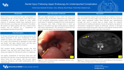
Case Description/Methods: An 86-year-old female with no prior gastrointestinal (GI) history presented to the clinic complaining of recent weight loss, fatigue, poor appetite, nausea, and vomiting for a week. She complained of dysphagia to both solids and liquids. In particular, she had trouble swallowing solid foods including meat and chicken. Vital signs, physical exam, and laboratory values were unremarkable and unchanged from baseline. EGD revealed benign esophageal stenosis and mild Schatzki’s ring, and both were dilated (Figure 1). The biopsy was negative for Helicobacter pylori or malignancy. The patient complained of new-onset tooth pain and discomfort the next day. It was revealed that the maxillary lateral incisor #11 was broken, with the gum above appearing edematous, and mildly erythematous with a retained tooth. WBC was slightly elevated at 13.9 × 10⁹/L. The chest X-ray revealed no signs of aspiration. Abdominal X-ray revealed an 8 mm nonspecific hyperdensity possibly within the distal small bowel. CT abdomen revealed 8-9 mm round density in the transverse colon, likely a foreign body or tooth cap (Figure 2). The patient was given broad-spectrum antibiotics with clinical improvement. The remainder of tooth #11 was extracted and the patient was discharged home in stable condition.
Discussion: The most frequently injured teeth during EGD are maxillary incisors, as they are thinner and have single roots compared to the more posterior teeth. When swallowed, broken teeth typically pass spontaneously through the stool in several days to weeks. Given our patient’s improvement in swelling and absence of acute GI symptoms, we opted for conservative management instead of endoscopic retrieval. A thorough physical exam noting loose teeth, dentures, and implants should be done preoperatively before an EGD, especially in the elderly.

Disclosures:
Farhan Azad, DO, Alexander M. Carlson, DO, Clive J. Miranda, DO, MS, Ross R. Moyer, DO, Prutha Patel, DO, Muddasir Ayaz, MD. P2016 - Dental Injury Following Upper Endoscopy: An Underreported Complication, ACG 2023 Annual Scientific Meeting Abstracts. Vancouver, BC, Canada: American College of Gastroenterology.
University at Buffalo, Buffalo, NY
Introduction: Dental injury is an uncommonly cited complication in endoscopy journals. At some centers, up to 40% of case cancellations can be seen related to teeth safety concerns. In anesthesia, dental injury represents up to half of all claims, occurring more commonly in the elderly. We present an elderly patient who had a lateral incisor avulsion and an infection as a complication of esophagogastroduodenoscopy (EGD).
Case Description/Methods: An 86-year-old female with no prior gastrointestinal (GI) history presented to the clinic complaining of recent weight loss, fatigue, poor appetite, nausea, and vomiting for a week. She complained of dysphagia to both solids and liquids. In particular, she had trouble swallowing solid foods including meat and chicken. Vital signs, physical exam, and laboratory values were unremarkable and unchanged from baseline. EGD revealed benign esophageal stenosis and mild Schatzki’s ring, and both were dilated (Figure 1). The biopsy was negative for Helicobacter pylori or malignancy. The patient complained of new-onset tooth pain and discomfort the next day. It was revealed that the maxillary lateral incisor #11 was broken, with the gum above appearing edematous, and mildly erythematous with a retained tooth. WBC was slightly elevated at 13.9 × 10⁹/L. The chest X-ray revealed no signs of aspiration. Abdominal X-ray revealed an 8 mm nonspecific hyperdensity possibly within the distal small bowel. CT abdomen revealed 8-9 mm round density in the transverse colon, likely a foreign body or tooth cap (Figure 2). The patient was given broad-spectrum antibiotics with clinical improvement. The remainder of tooth #11 was extracted and the patient was discharged home in stable condition.
Discussion: The most frequently injured teeth during EGD are maxillary incisors, as they are thinner and have single roots compared to the more posterior teeth. When swallowed, broken teeth typically pass spontaneously through the stool in several days to weeks. Given our patient’s improvement in swelling and absence of acute GI symptoms, we opted for conservative management instead of endoscopic retrieval. A thorough physical exam noting loose teeth, dentures, and implants should be done preoperatively before an EGD, especially in the elderly.

Figure: Figure 1: EGD showing benign esophageal stenosis and mild Schatzki’s ring (arrow)
Figure 2: CT abdomen showing an 8-9 mm round density in the transverse colon, likely a foreign body or tooth cap (arrow)
Figure 2: CT abdomen showing an 8-9 mm round density in the transverse colon, likely a foreign body or tooth cap (arrow)
Disclosures:
Farhan Azad indicated no relevant financial relationships.
Alexander Carlson indicated no relevant financial relationships.
Clive Miranda indicated no relevant financial relationships.
Ross Moyer indicated no relevant financial relationships.
Prutha Patel indicated no relevant financial relationships.
Muddasir Ayaz indicated no relevant financial relationships.
Farhan Azad, DO, Alexander M. Carlson, DO, Clive J. Miranda, DO, MS, Ross R. Moyer, DO, Prutha Patel, DO, Muddasir Ayaz, MD. P2016 - Dental Injury Following Upper Endoscopy: An Underreported Complication, ACG 2023 Annual Scientific Meeting Abstracts. Vancouver, BC, Canada: American College of Gastroenterology.
