Monday Poster Session
Category: GI Bleeding
P2056 - Average Risk Screening Colonoscopy Complicated by Intramural Hematoma and Hemoperitoneum in a Patient Without Any Risk Factor
Monday, October 23, 2023
10:30 AM - 4:15 PM PT
Location: Exhibit Hall

Has Audio
- ZL
Zilan Lin, MD
Westchester Medical Center
Valhalla, NY
Presenting Author(s)
Zilan Lin, MD, Alexander Levstik, MD, Agnieszka Nagorna, MD, Virendra Tewari, MD
Westchester Medical Center, Valhalla, NY
Introduction: Intramural hematoma is an extremely rare complication after colonoscopy and almost exclusively reported in cases with some risk factors such as anticoagulation, polypectomy, malignancy, etc. We report an unusual case of spontaneous intramural hematoma and hemoperitoneum in a patient without risk factors following an uneventful screening colonoscopy.
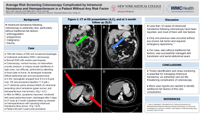
Case Description/Methods: A 74-year-old male underwent an elective outpatient esophagogastroduodenoscopy (EGD) and colonoscopy due to iron deficiency anemia, dysphagia, and colon cancer screening. His previous colonoscopy over 10 years ago was unremarkable. Random pan-biopsies were taken during his EGD. Retroflexion was performed in the right colon, and no other additional intervention was done during the colonoscopy. Abdominal counter pressure was not utilized. Colonic mucosa appeared to be normal. The procedure was performed by an experienced endoscopist. A few hours after returning home, the patient developed worsening abdominal pain, and syncopized twice, causing him to return to the hospital. Over 12 hours, his hemoglobin (Hb) declined from 9.5 to 6.8 (baseline Hb 11.5 g/dl). CT showed a 9cm intramural hematoma in the ascending colon, resulting in luminal narrowing, and a moderate hemoperitoneum (Figure 1 A,C). The patient was transfused with 2 units packed red blood cells, and his symptoms improved. He was monitored with serial abdominal exam, and Hb remained stable around 9 g/dl. He was discharged after 3 days. At 2 week outpatient follow up, Hb returned to baseline. CT 2 months later showed no hemoperitoneum, and a resolving hematoma from 9 to 5cm (Figure 1 B,D). No coagulopathy was noted by further work up.
Discussion: Spontaneous intramural hematoma after colonoscopy is highly uncommon. Very few case reports were previously published, less than 10 upon literature review. Most such cases had risk factors including anticoagulation, malignancy, or polypectomy. Apart from our patient, only one reported case was without any known risk factor.1 While our patient was successfully managed conservatively, the other case necessitated surgery. Requiring awareness, timely identification and close monitoring is essential for managing this extremely rare but life threatening complication of colonoscopy.
Reference:
1. Jalal A, Khatiwada P, Shoreibah M, et al. Intramural Hematoma and Retroperitoneal Hematoma Following a Routine Colonoscopy: An Uncommon Complication of a Common Procedure. Case Reports in Gastroenterology (May-Aug 2022) 16(2): 515-520.

Disclosures:
Zilan Lin, MD, Alexander Levstik, MD, Agnieszka Nagorna, MD, Virendra Tewari, MD. P2056 - Average Risk Screening Colonoscopy Complicated by Intramural Hematoma and Hemoperitoneum in a Patient Without Any Risk Factor, ACG 2023 Annual Scientific Meeting Abstracts. Vancouver, BC, Canada: American College of Gastroenterology.
Westchester Medical Center, Valhalla, NY
Introduction: Intramural hematoma is an extremely rare complication after colonoscopy and almost exclusively reported in cases with some risk factors such as anticoagulation, polypectomy, malignancy, etc. We report an unusual case of spontaneous intramural hematoma and hemoperitoneum in a patient without risk factors following an uneventful screening colonoscopy.
Case Description/Methods: A 74-year-old male underwent an elective outpatient esophagogastroduodenoscopy (EGD) and colonoscopy due to iron deficiency anemia, dysphagia, and colon cancer screening. His previous colonoscopy over 10 years ago was unremarkable. Random pan-biopsies were taken during his EGD. Retroflexion was performed in the right colon, and no other additional intervention was done during the colonoscopy. Abdominal counter pressure was not utilized. Colonic mucosa appeared to be normal. The procedure was performed by an experienced endoscopist. A few hours after returning home, the patient developed worsening abdominal pain, and syncopized twice, causing him to return to the hospital. Over 12 hours, his hemoglobin (Hb) declined from 9.5 to 6.8 (baseline Hb 11.5 g/dl). CT showed a 9cm intramural hematoma in the ascending colon, resulting in luminal narrowing, and a moderate hemoperitoneum (Figure 1 A,C). The patient was transfused with 2 units packed red blood cells, and his symptoms improved. He was monitored with serial abdominal exam, and Hb remained stable around 9 g/dl. He was discharged after 3 days. At 2 week outpatient follow up, Hb returned to baseline. CT 2 months later showed no hemoperitoneum, and a resolving hematoma from 9 to 5cm (Figure 1 B,D). No coagulopathy was noted by further work up.
Discussion: Spontaneous intramural hematoma after colonoscopy is highly uncommon. Very few case reports were previously published, less than 10 upon literature review. Most such cases had risk factors including anticoagulation, malignancy, or polypectomy. Apart from our patient, only one reported case was without any known risk factor.1 While our patient was successfully managed conservatively, the other case necessitated surgery. Requiring awareness, timely identification and close monitoring is essential for managing this extremely rare but life threatening complication of colonoscopy.
Reference:
1. Jalal A, Khatiwada P, Shoreibah M, et al. Intramural Hematoma and Retroperitoneal Hematoma Following a Routine Colonoscopy: An Uncommon Complication of a Common Procedure. Case Reports in Gastroenterology (May-Aug 2022) 16(2): 515-520.

Figure: Figure 1. CT findings at initial visit and at 2 month follow up. A. An intramural hematoma of 9.16cm at initial presentation. B. An intramural hematoma of 5.01cm at 2 month follow up. C. The presence of a moderate hemoperitoneum at initial presentation (arrows). D. Complete resolution of the hemoperitoneum at 2 month follow up.
Disclosures:
Zilan Lin indicated no relevant financial relationships.
Alexander Levstik indicated no relevant financial relationships.
Agnieszka Nagorna indicated no relevant financial relationships.
Virendra Tewari indicated no relevant financial relationships.
Zilan Lin, MD, Alexander Levstik, MD, Agnieszka Nagorna, MD, Virendra Tewari, MD. P2056 - Average Risk Screening Colonoscopy Complicated by Intramural Hematoma and Hemoperitoneum in a Patient Without Any Risk Factor, ACG 2023 Annual Scientific Meeting Abstracts. Vancouver, BC, Canada: American College of Gastroenterology.
