Monday Poster Session
Category: GI Bleeding
P2058 - Esophageal Dieulafoy: A Rare Cause of Gastrointestinal Hemorrhage in a Patient With SLE
Monday, October 23, 2023
10:30 AM - 4:15 PM PT
Location: Exhibit Hall

Has Audio

Abhishek A. Chouthai, MD
Cooper University Hospital at Cooper Medical School at Rowan University
Camden, NJ
Presenting Author(s)
Abhishek A. Chouthai, MD1, Eitan Scheinthal, DO1, Michael Schwartz, MD2, Cristina Capanescu, MD3
1Cooper University Hospital at Cooper Medical School at Rowan University, Camden, NJ; 2Cooper University Hospital, Camden, NJ; 3Digestive Health Institute at Cooper University Hospital, Camden, NJ
Introduction: A rare though potentially fatal cause of gastrointestinal bleeding, esophageal dieulafoy lesions are seldom identified. We report an esophageal dieulafoy in a patient with systemic lupus erythematosus (SLE) who was found to have massive hematemesis following nasogastric tube placement for small bowel obstruction.
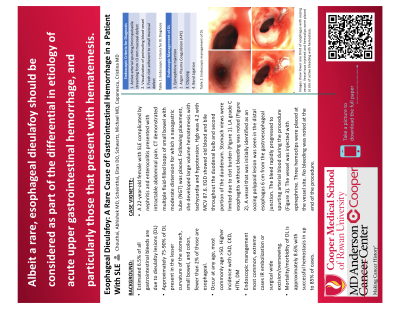
Case Description/Methods: A 22-year-old female with SLE complicated by nephritis and enterocolitis presented with intractable abdominal pain. CT demonstrated multiple fluid-filled loops of small bowel with moderate distension for which a nasogastric tube was placed. Following placement she developed large volume hematemesis. Hgb was 4.2 with MCV 87.6. EGD showed old blood and bile throughout the duodenal bulb and second portion of the duodenum. Views of the stomach were limited due to clot burden. LA grade C esophagitis without bleeding was noted. A vessel that was initially identified as an oozing polypoid lesion was seen in the distal esophagus 6 cm from the gastroesophageal junction. The bleed rapidly progressed to spurting arterial blood during the procedure. The vessel was injected with epinephrine and three hemoclips were placed at the vessel site. No bleeding was noted at the end of the procedure.
Discussion: An estimated 6.5% of all gastrointestinal bleeds are due to dieulafoy lesions, of which 2% are esophageal in origin. DL is elusive given its acute, intermittent pattern of bleeding and difficulty visualizing on endoscopy within typically normal appearing mucosa. Due to high risk of re-bleeding, DL biopsy is not recommended. Postulated mechanisms include large artery pulsation resulting in mechanical followed by chemical erosion, ischemia, vasculitis, and age related mucosal atrophy; NSAIDs, tobacco, alcohol, and peptic ulcer disease have previously been studied in correlation to vessel rupture but have no demonstrated association. While reported in all age groups, DL are most common in the elderly, particularly those with cardiovascular disease, hypertension, kidney disease and diabetes mellitus. Today, multimodal approaches for treatment include endoscopic clipping, epinephrine injection, thermocoagulation, or angiography with embolization and or wedge excision or oversewing may be utilized. With advances in endoscopy, hemostasis achieved in up to 85% of cases. Albeit a rare etiology, esophageal DL should be considered as part of the differential in etiology of acute upper gastrointestinal hemorrhage, and particularly those that present with hematemesis.
Disclosures:
Abhishek A. Chouthai, MD1, Eitan Scheinthal, DO1, Michael Schwartz, MD2, Cristina Capanescu, MD3. P2058 - Esophageal Dieulafoy: A Rare Cause of Gastrointestinal Hemorrhage in a Patient With SLE, ACG 2023 Annual Scientific Meeting Abstracts. Vancouver, BC, Canada: American College of Gastroenterology.
1Cooper University Hospital at Cooper Medical School at Rowan University, Camden, NJ; 2Cooper University Hospital, Camden, NJ; 3Digestive Health Institute at Cooper University Hospital, Camden, NJ
Introduction: A rare though potentially fatal cause of gastrointestinal bleeding, esophageal dieulafoy lesions are seldom identified. We report an esophageal dieulafoy in a patient with systemic lupus erythematosus (SLE) who was found to have massive hematemesis following nasogastric tube placement for small bowel obstruction.
Case Description/Methods: A 22-year-old female with SLE complicated by nephritis and enterocolitis presented with intractable abdominal pain. CT demonstrated multiple fluid-filled loops of small bowel with moderate distension for which a nasogastric tube was placed. Following placement she developed large volume hematemesis. Hgb was 4.2 with MCV 87.6. EGD showed old blood and bile throughout the duodenal bulb and second portion of the duodenum. Views of the stomach were limited due to clot burden. LA grade C esophagitis without bleeding was noted. A vessel that was initially identified as an oozing polypoid lesion was seen in the distal esophagus 6 cm from the gastroesophageal junction. The bleed rapidly progressed to spurting arterial blood during the procedure. The vessel was injected with epinephrine and three hemoclips were placed at the vessel site. No bleeding was noted at the end of the procedure.
Discussion: An estimated 6.5% of all gastrointestinal bleeds are due to dieulafoy lesions, of which 2% are esophageal in origin. DL is elusive given its acute, intermittent pattern of bleeding and difficulty visualizing on endoscopy within typically normal appearing mucosa. Due to high risk of re-bleeding, DL biopsy is not recommended. Postulated mechanisms include large artery pulsation resulting in mechanical followed by chemical erosion, ischemia, vasculitis, and age related mucosal atrophy; NSAIDs, tobacco, alcohol, and peptic ulcer disease have previously been studied in correlation to vessel rupture but have no demonstrated association. While reported in all age groups, DL are most common in the elderly, particularly those with cardiovascular disease, hypertension, kidney disease and diabetes mellitus. Today, multimodal approaches for treatment include endoscopic clipping, epinephrine injection, thermocoagulation, or angiography with embolization and or wedge excision or oversewing may be utilized. With advances in endoscopy, hemostasis achieved in up to 85% of cases. Albeit a rare etiology, esophageal DL should be considered as part of the differential in etiology of acute upper gastrointestinal hemorrhage, and particularly those that present with hematemesis.
Disclosures:
Abhishek Chouthai indicated no relevant financial relationships.
Eitan Scheinthal indicated no relevant financial relationships.
Michael Schwartz indicated no relevant financial relationships.
Cristina Capanescu indicated no relevant financial relationships.
Abhishek A. Chouthai, MD1, Eitan Scheinthal, DO1, Michael Schwartz, MD2, Cristina Capanescu, MD3. P2058 - Esophageal Dieulafoy: A Rare Cause of Gastrointestinal Hemorrhage in a Patient With SLE, ACG 2023 Annual Scientific Meeting Abstracts. Vancouver, BC, Canada: American College of Gastroenterology.
