Monday Poster Session
Category: GI Bleeding
P2078 - Unveiling the Hidden Serpent: Detecting a Non-Bleeding Colonic Dieulafoy’s Lesion After Positive FIT
Monday, October 23, 2023
10:30 AM - 4:15 PM PT
Location: Exhibit Hall

Has Audio
- JY
Jin Yun, DO
Providence Portland Medical Center
Portland, OR
Presenting Author(s)
Jin Yun, DO1, Branden Tarlow, MD2
1Providence Portland Medical Center, Portland, OR; 2The Oregon Clinic, Portland, OR
Introduction: Dieulafoy’s lesions are an uncommon but important cause of intermittent gastrointestinal bleeding as they can be life-threatening, in part due to delayed diagnosis. Published studies propose Dieulafoy’s lesions account for 1-2% of acute gastrointestinal bleeds, but their true prevalence may be underestimated due to a lack of recognition and confirmation. We report a case of a non-bleeding Dieulafoy’s lesion detected in the cecum with optical diagnosis technique and histologic correlation.
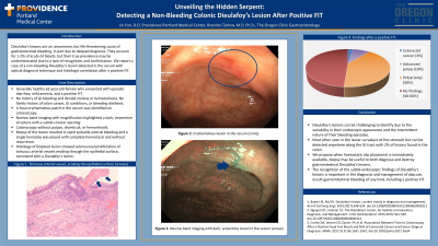
Case Description/Methods: A 62-year-old woman with a history of hypertension presented to the outpatient endoscopy center with episodic diarrhea, mild anemia, and a positive fecal immunochemical test (FIT). She had no history of gastrointestinal bleeding and denied melena or hematochezia. She did not have a family history of colon cancer, gastrointestinal conditions, or bleeding diathesis. Her physical exam was unremarkable. The patient underwent a colonoscopy with identification of a focal erythematous patch in the cecum. Narrow band imaging with magnification highlighted a dark, serpentine structure with subtle circular opening. Biopsy of the lesion resulted in rapid pulsatile arterial bleeding and a single hemoclip was placed with complete hemostasis and without recurrence. The exam was otherwise notable for an absence of polyps, diverticuli, or hemorrhoids. Histology of the biopsied lesion revealed submucosal proliferation of tortuous arterial vessels eroding through the epithelial surface without ulceration, consistent with a Dieulafoy’s lesion.
Discussion: Dieulafoy’s lesions can be challenging to identify due to the variability in their endoscopic appearance and the intermittent nature of their bleeding episodes. They are most often seen in the lesser curvature of the stomach, but can be detected anywhere along the gastrointestinal tract with 2% of lesions found in the colon. We propose when hemostatic clip placement is immediately available, biopsy may be useful to both diagnose and destroy gastrointestinal Dieulafoy’s lesions. Therapeutic endoscopy using hemostatic clips have been studied to be safe and highly effective treatment for Dieulafoy’s lesions and are thought to offer similar hemostatic efficacy as combination therapy in better initial hemostasis and rebleeding outcomes. We conclude the recognition of the subtle endoscopic findings of Dieulafoy’s lesions is important in the diagnosis and management of obscure, occult gastrointestinal bleeding of any kind, including a positive FIT.
Disclosures:
Jin Yun, DO1, Branden Tarlow, MD2. P2078 - Unveiling the Hidden Serpent: Detecting a Non-Bleeding Colonic Dieulafoy’s Lesion After Positive FIT, ACG 2023 Annual Scientific Meeting Abstracts. Vancouver, BC, Canada: American College of Gastroenterology.
1Providence Portland Medical Center, Portland, OR; 2The Oregon Clinic, Portland, OR
Introduction: Dieulafoy’s lesions are an uncommon but important cause of intermittent gastrointestinal bleeding as they can be life-threatening, in part due to delayed diagnosis. Published studies propose Dieulafoy’s lesions account for 1-2% of acute gastrointestinal bleeds, but their true prevalence may be underestimated due to a lack of recognition and confirmation. We report a case of a non-bleeding Dieulafoy’s lesion detected in the cecum with optical diagnosis technique and histologic correlation.
Case Description/Methods: A 62-year-old woman with a history of hypertension presented to the outpatient endoscopy center with episodic diarrhea, mild anemia, and a positive fecal immunochemical test (FIT). She had no history of gastrointestinal bleeding and denied melena or hematochezia. She did not have a family history of colon cancer, gastrointestinal conditions, or bleeding diathesis. Her physical exam was unremarkable. The patient underwent a colonoscopy with identification of a focal erythematous patch in the cecum. Narrow band imaging with magnification highlighted a dark, serpentine structure with subtle circular opening. Biopsy of the lesion resulted in rapid pulsatile arterial bleeding and a single hemoclip was placed with complete hemostasis and without recurrence. The exam was otherwise notable for an absence of polyps, diverticuli, or hemorrhoids. Histology of the biopsied lesion revealed submucosal proliferation of tortuous arterial vessels eroding through the epithelial surface without ulceration, consistent with a Dieulafoy’s lesion.
Discussion: Dieulafoy’s lesions can be challenging to identify due to the variability in their endoscopic appearance and the intermittent nature of their bleeding episodes. They are most often seen in the lesser curvature of the stomach, but can be detected anywhere along the gastrointestinal tract with 2% of lesions found in the colon. We propose when hemostatic clip placement is immediately available, biopsy may be useful to both diagnose and destroy gastrointestinal Dieulafoy’s lesions. Therapeutic endoscopy using hemostatic clips have been studied to be safe and highly effective treatment for Dieulafoy’s lesions and are thought to offer similar hemostatic efficacy as combination therapy in better initial hemostasis and rebleeding outcomes. We conclude the recognition of the subtle endoscopic findings of Dieulafoy’s lesions is important in the diagnosis and management of obscure, occult gastrointestinal bleeding of any kind, including a positive FIT.
Disclosures:
Jin Yun indicated no relevant financial relationships.
Branden Tarlow indicated no relevant financial relationships.
Jin Yun, DO1, Branden Tarlow, MD2. P2078 - Unveiling the Hidden Serpent: Detecting a Non-Bleeding Colonic Dieulafoy’s Lesion After Positive FIT, ACG 2023 Annual Scientific Meeting Abstracts. Vancouver, BC, Canada: American College of Gastroenterology.
