Monday Poster Session
Category: GI Bleeding
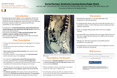
P2089 - Buried Bumper Syndrome Leading to Hemorrhagic Shock
Monday, October 23, 2023
10:30 AM - 4:15 PM PT
Location: Exhibit Hall

Has Audio

Amit Sah, MD
University of Miami at JFK Medical Center
Lantana, FL
Presenting Author(s)
Amit Sah, MD1, Lesley- Ann McCook, MD2, Elizabeth Zakka, MD1, Akiva Marcus, MD3, Kayode Olowe, MD4
1University of Miami at JFK Medical Center, Lantana, FL; 2University of Miami, Atlantis, FL; 3HCA Florida Physicians, JFK Medical Center, West Palm Beach, FL; 4HCA Healthcare, Atlantis, FL
Introduction: Buried Bumper Syndrome (BBS) is a rare complication of PEG tube due to the migration of internal fixation devices alongside the track of the stoma. It is reported in about 1% of patients with PEG tube, traditionally as a late complication. It usually occurs in immunocompromised, malnourished with poor wound healing and most importantly with high tension between internal and external bolsters. The common manifestation of BBS is peristomal leakage, local infection, inability to insert content, etc. Here we present a case of BBS causing hemorrhagic shock.
Case Description/Methods: A 55-year-old male presented with chronic dysphagia of 2-3 months duration. He had normocytic anemia with Hgb 11.2 g/dL and AKI on presentation. Video swallow showed tracheal aspiration with unable to clear pharyngeal residues. During the evaluation, he was diagnosed with dermatomyositis which is likely the cause of his dysphagia. He underwent a laparoscopic-assisted PEG tube placement. Two days postoperatively, he developed hemorrhagic shock. After stabilization, he underwent EGD which was limited due to poor visualization from blood products throughout the stomach. The PEG tube was not visualized in the stomach and there was transillumination on the epigastric region at the site of the PEG tube.
CT abdomen and pelvis with contrast infusion through PEG tube demonstrated PEG tube tip outside the stomach and extravasated contrast within the peritoneum overlying the stomach and liver. He underwent an IR guided angiogram that demonstrated an active extravasation from the right gastric artery which was successfully embolized. TPN was started for nutrition and he later had surgical creation of a PEG tube at a different site. He continued to improve and after an extensive clinical course and with the help of the multidisciplinary team, he was discharged to home with family.
Discussion: BBS is a rare and traditionally a late complication of PEG tube placement. It is complicated with abscess, pain, and peritonitis, and rarely with UGIB. It is classified as Grade I, II, and III referring to partial, subtotal, and total migration respectively. Grade I is managed endoscopically while II and III are managed surgically. Our patient's BBS was grade II complicated with severe UGIB and hence he later had a surgical PEG tube placement at a different site. Although rare, PEG tube complications of early BBS should be considered in the case of UGIB. A multidisciplinary and timely evaluation and management is the key to a better outcome.

Disclosures:
Amit Sah, MD1, Lesley- Ann McCook, MD2, Elizabeth Zakka, MD1, Akiva Marcus, MD3, Kayode Olowe, MD4. P2089 - Buried Bumper Syndrome Leading to Hemorrhagic Shock, ACG 2023 Annual Scientific Meeting Abstracts. Vancouver, BC, Canada: American College of Gastroenterology.
1University of Miami at JFK Medical Center, Lantana, FL; 2University of Miami, Atlantis, FL; 3HCA Florida Physicians, JFK Medical Center, West Palm Beach, FL; 4HCA Healthcare, Atlantis, FL
Introduction: Buried Bumper Syndrome (BBS) is a rare complication of PEG tube due to the migration of internal fixation devices alongside the track of the stoma. It is reported in about 1% of patients with PEG tube, traditionally as a late complication. It usually occurs in immunocompromised, malnourished with poor wound healing and most importantly with high tension between internal and external bolsters. The common manifestation of BBS is peristomal leakage, local infection, inability to insert content, etc. Here we present a case of BBS causing hemorrhagic shock.
Case Description/Methods: A 55-year-old male presented with chronic dysphagia of 2-3 months duration. He had normocytic anemia with Hgb 11.2 g/dL and AKI on presentation. Video swallow showed tracheal aspiration with unable to clear pharyngeal residues. During the evaluation, he was diagnosed with dermatomyositis which is likely the cause of his dysphagia. He underwent a laparoscopic-assisted PEG tube placement. Two days postoperatively, he developed hemorrhagic shock. After stabilization, he underwent EGD which was limited due to poor visualization from blood products throughout the stomach. The PEG tube was not visualized in the stomach and there was transillumination on the epigastric region at the site of the PEG tube.
CT abdomen and pelvis with contrast infusion through PEG tube demonstrated PEG tube tip outside the stomach and extravasated contrast within the peritoneum overlying the stomach and liver. He underwent an IR guided angiogram that demonstrated an active extravasation from the right gastric artery which was successfully embolized. TPN was started for nutrition and he later had surgical creation of a PEG tube at a different site. He continued to improve and after an extensive clinical course and with the help of the multidisciplinary team, he was discharged to home with family.
Discussion: BBS is a rare and traditionally a late complication of PEG tube placement. It is complicated with abscess, pain, and peritonitis, and rarely with UGIB. It is classified as Grade I, II, and III referring to partial, subtotal, and total migration respectively. Grade I is managed endoscopically while II and III are managed surgically. Our patient's BBS was grade II complicated with severe UGIB and hence he later had a surgical PEG tube placement at a different site. Although rare, PEG tube complications of early BBS should be considered in the case of UGIB. A multidisciplinary and timely evaluation and management is the key to a better outcome.

Figure: CT scan of the Abdomen and Pelvis with Contrast via PEG tube showing extravasation within the peritoneum and the PEG tube tip outside the stomach.
Disclosures:
Amit Sah indicated no relevant financial relationships.
Lesley- Ann McCook indicated no relevant financial relationships.
Elizabeth Zakka indicated no relevant financial relationships.
Akiva Marcus indicated no relevant financial relationships.
Kayode Olowe indicated no relevant financial relationships.
Amit Sah, MD1, Lesley- Ann McCook, MD2, Elizabeth Zakka, MD1, Akiva Marcus, MD3, Kayode Olowe, MD4. P2089 - Buried Bumper Syndrome Leading to Hemorrhagic Shock, ACG 2023 Annual Scientific Meeting Abstracts. Vancouver, BC, Canada: American College of Gastroenterology.
