Monday Poster Session
Category: Interventional Endoscopy
P2325 - Gallstone Removal Resulting in Resolution of a Rare Case of Bouveret Syndrome
Monday, October 23, 2023
10:30 AM - 4:15 PM PT
Location: Exhibit Hall

Has Audio

Charlie Altfillisch, MD
University of Kansas Medical Center
Kansas City, KS
Presenting Author(s)
Charlie Altfillisch, MD1, Matthew Moore, MD2, Mojtaba Olyaee, MD2
1University of Kansas Medical Center, Kansas City, KS; 2University of Kansas, Kansas City, KS
Introduction: Bouveret syndrome is a rare variant of gallstone ileus caused by an acquired fistula via the gallbladder and either the stomach or duodenum. Fistulization occurs due to inflammation and pressure via gallstones following acute cholecystitis which may result in an ischemic tear. There are approximately 300 documented case reports over the past 50 years, as Bouveret syndrome accounts for 1-3% of cases of gallstone ileus and affects < 0.5% of patients with gallstones. The nonspecific symptoms make diagnosis difficult, as the majority of patients present with nausea, emesis and abdominal pain.
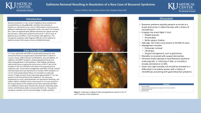
Case Description/Methods: A 77-year-old female with PMH of complicated diverticulitis with colovesical fistula s/p surgical repair presented with 2-3 weeks of nausea, emesis, abdominal pain and distention, and constipation. CT abdomen and MRCP revealed a cholecystoduodenal fistula and obstructing gallstone in the duodenum. EGD findings included an obstructing stone occupying the entire lumen of the 3rd part of the duodenum and circumferential ulcerations from presumed stone impaction sites. Conservative management with NPO, IV fluids, and NG tube decompression was started prior to transfer. ERCP revealed a 12-15 mm cholecystoduodenal fistula not amenable to endoscopic closure. A large occlusive stone measuring approximately 5-7 cm was present in the third portion of the duodenum. The stone was fragmented via snare, electrohydraulic and mechanical lithotripsy, and balloon dilation. Multiple stones measuring 8-15 mm were present in the second portion of the duodenum and were extracted with Roth net. Additional stones were moved to the stomach and broken down further with lithotripsy before removal with Roth net. The patient's symptoms resolved, and she was discharged in stable condition.
Discussion: Bouveret syndrome typically presents as similar to a bowel obstruction in elderly females with a history of cholelithiasis. Imaging may reveal Rigler’s triad (dilated stomach, pneumobilia, radio-opaque shadow), although, this triad is only present in 40-50% of cases. Management includes endoscopic removal, lithotripsy, or surgical management such as gastrotomy. Mortality is increased with surgical intervention and clinicians should attempt to treat Bouveret syndrome endoscopically +/- lithotripsy if able, as mortality is already estimated at 12-30%. Given such high mortality, this should be included on a differential in an elderly woman with a history of cholelithiasis presenting with gastrointestinal symptoms.

Disclosures:
Charlie Altfillisch, MD1, Matthew Moore, MD2, Mojtaba Olyaee, MD2. P2325 - Gallstone Removal Resulting in Resolution of a Rare Case of Bouveret Syndrome, ACG 2023 Annual Scientific Meeting Abstracts. Vancouver, BC, Canada: American College of Gastroenterology.
1University of Kansas Medical Center, Kansas City, KS; 2University of Kansas, Kansas City, KS
Introduction: Bouveret syndrome is a rare variant of gallstone ileus caused by an acquired fistula via the gallbladder and either the stomach or duodenum. Fistulization occurs due to inflammation and pressure via gallstones following acute cholecystitis which may result in an ischemic tear. There are approximately 300 documented case reports over the past 50 years, as Bouveret syndrome accounts for 1-3% of cases of gallstone ileus and affects < 0.5% of patients with gallstones. The nonspecific symptoms make diagnosis difficult, as the majority of patients present with nausea, emesis and abdominal pain.
Case Description/Methods: A 77-year-old female with PMH of complicated diverticulitis with colovesical fistula s/p surgical repair presented with 2-3 weeks of nausea, emesis, abdominal pain and distention, and constipation. CT abdomen and MRCP revealed a cholecystoduodenal fistula and obstructing gallstone in the duodenum. EGD findings included an obstructing stone occupying the entire lumen of the 3rd part of the duodenum and circumferential ulcerations from presumed stone impaction sites. Conservative management with NPO, IV fluids, and NG tube decompression was started prior to transfer. ERCP revealed a 12-15 mm cholecystoduodenal fistula not amenable to endoscopic closure. A large occlusive stone measuring approximately 5-7 cm was present in the third portion of the duodenum. The stone was fragmented via snare, electrohydraulic and mechanical lithotripsy, and balloon dilation. Multiple stones measuring 8-15 mm were present in the second portion of the duodenum and were extracted with Roth net. Additional stones were moved to the stomach and broken down further with lithotripsy before removal with Roth net. The patient's symptoms resolved, and she was discharged in stable condition.
Discussion: Bouveret syndrome typically presents as similar to a bowel obstruction in elderly females with a history of cholelithiasis. Imaging may reveal Rigler’s triad (dilated stomach, pneumobilia, radio-opaque shadow), although, this triad is only present in 40-50% of cases. Management includes endoscopic removal, lithotripsy, or surgical management such as gastrotomy. Mortality is increased with surgical intervention and clinicians should attempt to treat Bouveret syndrome endoscopically +/- lithotripsy if able, as mortality is already estimated at 12-30%. Given such high mortality, this should be included on a differential in an elderly woman with a history of cholelithiasis presenting with gastrointestinal symptoms.

Figure: Endoscopic imaging of obstructing gallstones present in 2nd and 3rd portions of the duodenum
Disclosures:
Charlie Altfillisch indicated no relevant financial relationships.
Matthew Moore indicated no relevant financial relationships.
Mojtaba Olyaee indicated no relevant financial relationships.
Charlie Altfillisch, MD1, Matthew Moore, MD2, Mojtaba Olyaee, MD2. P2325 - Gallstone Removal Resulting in Resolution of a Rare Case of Bouveret Syndrome, ACG 2023 Annual Scientific Meeting Abstracts. Vancouver, BC, Canada: American College of Gastroenterology.
