Monday Poster Session
Category: Liver
P2430 - Patterns of Abnormal MUC1 Expression in Patients with PSC and Cholangiocarcinoma
Monday, October 23, 2023
10:30 AM - 4:15 PM PT
Location: Exhibit Hall

Has Audio

Jana G. Al Hashash, MD, MSc, FACG
Associate Professor
Mayo Clinic
Jacksonville, FL
Presenting Author(s)
Award: Outstanding Research Award in the Liver Category
Award: Presidential Poster Award
Pamela Beatty, PhD1, Irene Yan, 2, Fadi Francis, MD1, Raouf Nakhleh, MD2, Denise Harnois, DO3, Francis A. Farraye, MD, MSc, MACG2, Tushar Patel, MBChB2, Jana G.. Hashash, MD, MSc, FACG2
1University of Pittsburgh, Pittsburgh, PA; 2Mayo Clinic, Jacksonville, FL; 3Mayo Clinic Florida, Jacksonville, FL
Introduction: MUC1 glycoprotein is a mucin expressed at low levels and in fully glycosylated form on healthy epithelial cells. Inflammation and cancer result in MUC1 becoming overexpressed and hypoglycosylated. Our aim was to describe patterns of MUC1 expression in patients with primary sclerosing cholangitis (PSC), PSC/cholangiocarcinoma, sporadic cholangiocarcinoma, and healthy controls. We hypothesize that overexpression of hypoglycosylated MUC1 would be found in small to moderate amount on bile duct epithelial cells of patients with PSC, higher levels in patients with PSC and concomitant cholangiocarcinoma, and even higher levels in patients with sporadic cholangiocarcinoma.
Methods: Archived tissue from liver explants of patients with PSC, PSC/cholangiocarcinoma, and cholangiocarcinoma were identified. Controls included normal liver biopsy tissue. Consecutive tissue sections from the hila (except among the controls) were stained using two different anti-MUC1 antibodies; HMPV that recognizes all forms of MUC1 and 4H5 that only recognizes abnormal hypoglycosylated MUC1. Each slide was graded based on quantity of staining (score 0-4) and intensity (score 0-4) by a pathologist blinded to the clinical information. The average of these 2 values was used to calculate a total MUC1 expression score.
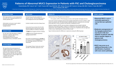
Results: Archived tissue from 38 patients were obtained: 5 controls, 22 PSC, 5 PSC/cholangiocarcinoma, and 6 sporadic cholangiocarcinoma. Tissue from controls minimally expressed the hypoglycosylated/abnormal form of MUC1, while tissue from patients with PSC demonstrated moderate expression compared to the very high levels expressed in tissue of patients with sporadic cholangiocarcinoma (Figure 1A). Explants of patients with PSC and concomitant cholangiocarcinoma showed moderate expression of the abnormal MUC1, however it is important to note that all 5 patients were treated with chemo/radiation therapy leading to scarring, loss of epithelial cells and regression of their cancer, interfering with accuracy of these results. Figure 1B demonstrates the difference in expression among the different groups.
Discussion: Abnormal MUC1 is not or minimally expressed in normal bile duct epithelial cells. Moderate overexpression of the hypoglycosylated form was found in bile duct epithelia of patients with PSC and to a greater degree in patients with cholangiocarcinoma. MUC1 may serve as an additional biomarker in the identification of patients with more severe pathology.

Disclosures:
Pamela Beatty, PhD1, Irene Yan, 2, Fadi Francis, MD1, Raouf Nakhleh, MD2, Denise Harnois, DO3, Francis A. Farraye, MD, MSc, MACG2, Tushar Patel, MBChB2, Jana G.. Hashash, MD, MSc, FACG2. P2430 - Patterns of Abnormal MUC1 Expression in Patients with PSC and Cholangiocarcinoma, ACG 2023 Annual Scientific Meeting Abstracts. Vancouver, BC, Canada: American College of Gastroenterology.
Award: Presidential Poster Award
Pamela Beatty, PhD1, Irene Yan, 2, Fadi Francis, MD1, Raouf Nakhleh, MD2, Denise Harnois, DO3, Francis A. Farraye, MD, MSc, MACG2, Tushar Patel, MBChB2, Jana G.. Hashash, MD, MSc, FACG2
1University of Pittsburgh, Pittsburgh, PA; 2Mayo Clinic, Jacksonville, FL; 3Mayo Clinic Florida, Jacksonville, FL
Introduction: MUC1 glycoprotein is a mucin expressed at low levels and in fully glycosylated form on healthy epithelial cells. Inflammation and cancer result in MUC1 becoming overexpressed and hypoglycosylated. Our aim was to describe patterns of MUC1 expression in patients with primary sclerosing cholangitis (PSC), PSC/cholangiocarcinoma, sporadic cholangiocarcinoma, and healthy controls. We hypothesize that overexpression of hypoglycosylated MUC1 would be found in small to moderate amount on bile duct epithelial cells of patients with PSC, higher levels in patients with PSC and concomitant cholangiocarcinoma, and even higher levels in patients with sporadic cholangiocarcinoma.
Methods: Archived tissue from liver explants of patients with PSC, PSC/cholangiocarcinoma, and cholangiocarcinoma were identified. Controls included normal liver biopsy tissue. Consecutive tissue sections from the hila (except among the controls) were stained using two different anti-MUC1 antibodies; HMPV that recognizes all forms of MUC1 and 4H5 that only recognizes abnormal hypoglycosylated MUC1. Each slide was graded based on quantity of staining (score 0-4) and intensity (score 0-4) by a pathologist blinded to the clinical information. The average of these 2 values was used to calculate a total MUC1 expression score.
Results: Archived tissue from 38 patients were obtained: 5 controls, 22 PSC, 5 PSC/cholangiocarcinoma, and 6 sporadic cholangiocarcinoma. Tissue from controls minimally expressed the hypoglycosylated/abnormal form of MUC1, while tissue from patients with PSC demonstrated moderate expression compared to the very high levels expressed in tissue of patients with sporadic cholangiocarcinoma (Figure 1A). Explants of patients with PSC and concomitant cholangiocarcinoma showed moderate expression of the abnormal MUC1, however it is important to note that all 5 patients were treated with chemo/radiation therapy leading to scarring, loss of epithelial cells and regression of their cancer, interfering with accuracy of these results. Figure 1B demonstrates the difference in expression among the different groups.
Discussion: Abnormal MUC1 is not or minimally expressed in normal bile duct epithelial cells. Moderate overexpression of the hypoglycosylated form was found in bile duct epithelia of patients with PSC and to a greater degree in patients with cholangiocarcinoma. MUC1 may serve as an additional biomarker in the identification of patients with more severe pathology.

Figure: Figure 1A demonstrates the staining patterns of abnormal MUC1 expression on bile ducts of normal controls, PSC, PSC/cholangiocarcinoma, and sporadic cholangiocarcinoma patients. Figure 1B is a bar graph showing the differences in abnormal MUC1 staining scores (as measured by 4H5 antibody) between the 4 study groups; each dot represents a single patient.
Disclosures:
Pamela Beatty indicated no relevant financial relationships.
Irene Yan indicated no relevant financial relationships.
Fadi Francis indicated no relevant financial relationships.
Raouf Nakhleh indicated no relevant financial relationships.
Denise Harnois indicated no relevant financial relationships.
Francis Farraye: AbbVie – Advisory Committee/Board Member. Avalo Therapeutics – Advisory Committee/Board Member. BMS – Advisory Committee/Board Member. Braintree Labs – Advisory Committee/Board Member. Fresenius Kabi – Advisory Committee/Board Member. GI Reviewers – Independent Contractor. GSK – Advisory Committee/Board Member. IBD Educational Group – Independent Contractor. Iterative Health – Advisory Committee/Board Member. Janssen – Advisory Committee/Board Member. Pfizer – Advisory Committee/Board Member. Pharmacosmos – Advisory Committee/Board Member. Sandoz Immunology – Advisory Committee/Board Member. Sebela – Advisory Committee/Board Member. Viatris – Advisory Committee/Board Member.
Tushar Patel indicated no relevant financial relationships.
Jana Hashash: Iterative Health – Grant/Research Support.
Pamela Beatty, PhD1, Irene Yan, 2, Fadi Francis, MD1, Raouf Nakhleh, MD2, Denise Harnois, DO3, Francis A. Farraye, MD, MSc, MACG2, Tushar Patel, MBChB2, Jana G.. Hashash, MD, MSc, FACG2. P2430 - Patterns of Abnormal MUC1 Expression in Patients with PSC and Cholangiocarcinoma, ACG 2023 Annual Scientific Meeting Abstracts. Vancouver, BC, Canada: American College of Gastroenterology.


